Introduction
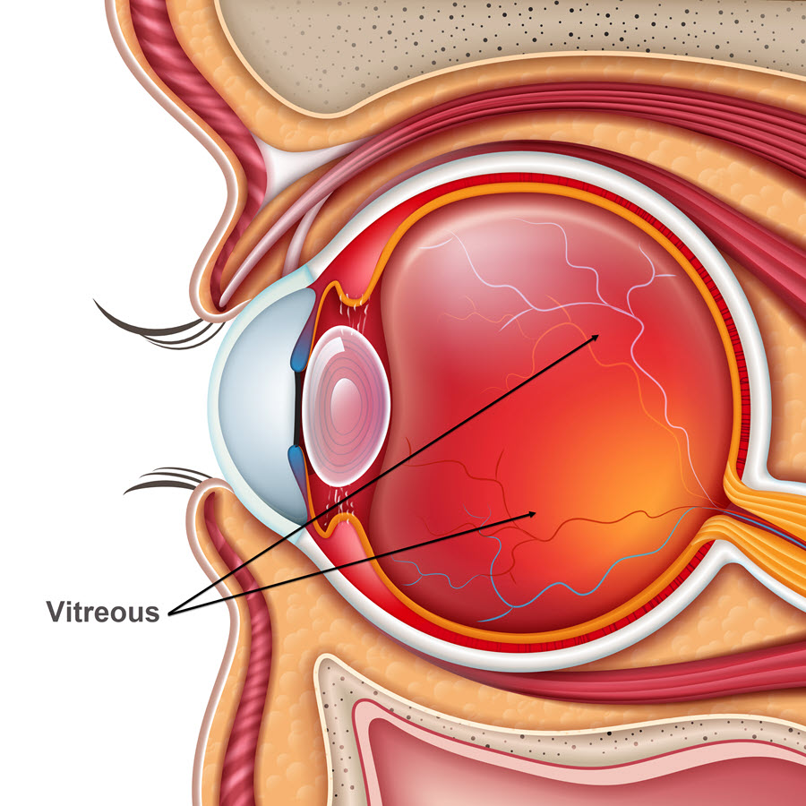
Between the lens and the retina lies the vitreous cavity which is filled with vitreous humor. The vitreous nourishes the inner parts of the eye. When light enters the eye, it passes through the vitreous on its way to the retina for projection.
Also Known As
- Hyaloid
- Vitreous humor
Anatomy
The vitreous occupies about 80% of the space inside the posterior of the eye. It is a jelly-like substance whose base is firmly attached to the retina. It is composed mainly of water (99%). Proteins, collagen and cells used to maintain clarity also form part of the vitreous. It is one of the most delicate connective body tissues. Because of the high water content, the vitreous chamber may look empty.
Function
The vitreous allows light to pass from the lens and retina enabling vision. It also maintains the eyeball’s spherical shape by its mass.
Associated symptoms & disorders
The following conditions are associated with the vitreous:
- Posterior vitreous detachment - Due to age, vitreous humor loosens, sometimes forcing it to tug on the retina. Posterior vitreous detachment occurs when the force of pulling is so strong that it separates the vitreous humor from the retina at the back of the eye. It can lead to loss of vision.
- Vitreous hemorrhage is a common cause of vision loss where blood clots form in the vitreous body. It is caused by proliferative diabetic retinopathy, ocular trauma and posterior vitreous detachment.
- Persistent hyperplastic primary vitreous or persistent fetal vasculature (PFV) occurs when the regression between the fetal hyaloid artery and the primary vitreous fails to take place. The result is an eye that is smaller than the other. The condition can lead to opacity (cataract) of the lens. It can also lead to angle-closure glaucoma.
- Synchysis scintillans is the degenerative liquefaction of the vitreous with cholesterol crystals accumulating in the vitreous. It may result from vitreous hemorrhage with the presence of floaters.
- Asteroid hyalosis (AH) occurs when lipid complexes are suspended throughout the collagen fibrils of the vitreous. It is associated with several systemic diseases as well as age. The disease rarely causes visual disturbance.
- Vitreomacular traction (VMT) syndrome refers to a disorder associated with the vitreoretinal interface. It usually features an incomplete PVD.
- Preretinal subhyaloid hemorrhage is a disease that leads to blood clots between the retina and the posterior vitreous membrane. It can result in a hyphema and black opacities or loss of vision.
Diagnosis of associated disorders
Some of the tests that are associated with the vitreous may include:
- A gonioscopy test
- A dilated fundus examination
- A slit-lamp examination to check structures at the front of the eye
- A B-scan ultranoscopy can detect vitreous hemorrhage
- Laboratory tests if diabetes is suspected
- Imaging tests such as computed tomography and magnetic resonance imaging
- An orbital CT scan to assess the integrity of the eyewall and rule out a foreign body
- A physical examination, for instance, to look for blood clots in the case of vitreous hemorrhage
- Indirect ophthalmoscopy can help diagnose posterior vitreous detachment. Others include echography to detect retinal tears
Treatment of associated disorders
Currently, there is no known medical treatment for conditions like posterior vitreous detachment. The first line of treatment is to put the patient under observation. However, different types of vitrectomy (removal of the vitreous) like pars plana, can assist if there is no change. Synchysis scintillans follows a similar treatment procedure.
For conditions like vitreous hemorrhage, it's best to treat the underlying medical disease such as diabetes with options such as Panretinal photocoagulation laser surgery and pars plana vitrectomy are options. Anti-VEGF drugs treat diabetic patients with vitreous hemorrhage. They also treat diabetic patients with neovascular age-related macular degeneration. Doctors advise a period of observation first before considering surgical treatment options.
Lensectomy, membranectomy and vitrectomy can treat PFV. Cataract surgery will help if the lens becomes opaque due to cataract(s). Other treatment options for AH include refraction and amblyopia therapy. Since glaucoma is a complication of PFV, the doctor administers glaucoma medications such as eye injections.
To manage VMT, patients can do a routine self-assessment using an Amsler grid evaluation. Researchers are still working on several medical therapies such as pharmacologic vitreolysis. Others undergoing research include enzymatic agents like jetrea, dispase, collagenase, chondroitinase, nattokinase and hyaluronidase.
Laser hyaloidotomy and pars plana vitrectomy can treat preretinal subhyaloid hemorrhage. In laser hyaloidotomy, the surgeon opens up the hyaloid membrane of the vitreous body and blood is distributed by diffusion.
