Introduction
Rods and cones are special cells situated at the back of the eye. They are generally referred to as photoreceptors. The cells convert light that enters into the eye into signals which are then sent to the brain.
Rod cells are found on the outer region of the retina. The cone cells, on the other hand, are densely packed in the fovea centralis. There are over 100 million rods and about 6 million cone cells in the human eye.
Anatomy
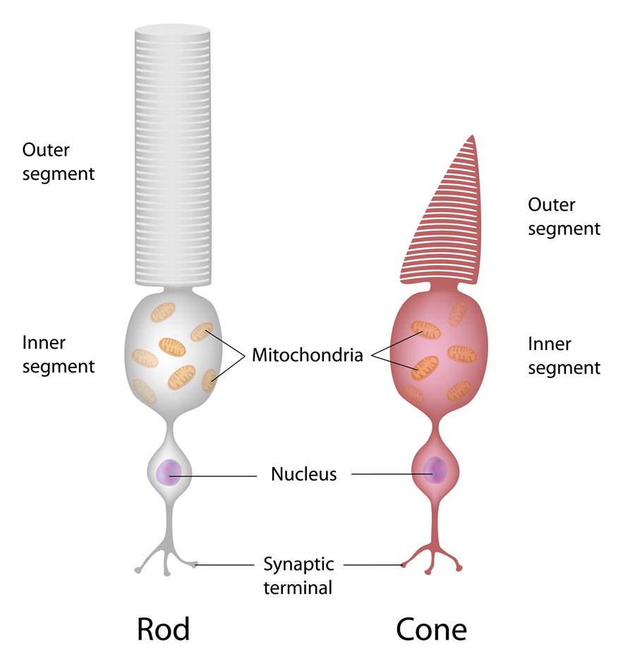
Rods and cones are quite similar in their structure. However, rods are more elongated and are narrower than the cones. They also have a rod-shaped end whereas cones have a cone shape at the end. Both cells have an outer segment, an inner segment and the synaptic area.
The outer segment contains the compact-packed disks where the photoreceptors molecules (rhodopsin for rods and iodopsin for cones) are located. The inner segment of the cells has the mitochondria, organelles and the cell nucleus. The synaptic region is the area where the cells relay information to intermediate neurons in the retina.
When light enters the cells, the proteins are converted to the proteins retinal and opsin (pigment). In cones, the pigments are of three types. They are:
- Blue-sensitive cones (S-cones) - These cells respond to light of short wavelengths. They have the highest sensitivity. They are found outside of the fovea centralis.
- Green-sensitive cones (M-cones) - The cones respond to light with medium wavelengths. They are densely packed in the fovea.
- Red-sensitive cones – They respond to light of long wavelengths. Together with the M-cones, they are packed at the center of the retina.
Function
Rods are responsible for night-time vision and peripheral vision. This is because they are highly sensitive to light and can therefore pick photons even in dim light. Being located on the outer region makes them proper for side vision. But they do not sense color and image details.
Cones are less sensitive to light but are tasked with color vision. They are active during the day when the light levels are high. Since they are centrally located, they also help in seeing fine details. When the three color pigments overlap, the brain interprets the signals and a person can see a variety of colors.
Associated symptoms & disorders
Diseases of the cones and rods occur when the cells are faulty, when they are damaged or when the path from the cell to the brain has a problem. The disorders can include:
- Color blindness - This is a condition where a person does not perceive color correctly. It happens when the cone cells do not work or are missing. There are three types of the condition. They include complete color blindness, red-green color blindness, and blue-yellow color blindness.
- Retinitis pigmentosa – It also referred to as rod-cone dystrophy. It is a condition of the loss of retinal cells. The rods are affected first and the cones follow. Onset symptoms include loss of side vision and night-time vision. With time, it becomes difficult to see fine details.
- Usher syndrome – The disease mostly occurs secondary to retinitis pigmentosa. It manifests as partial/complete loss of vision and hearing. As the photoreceptors gradually deteriorate, blind spots form which then produce tunnel vision.
- Photokeratitis – This is an eye condition that occurs when exposure to unfiltered UV rays damages the eye. In the retina, the intense light triggers chemical reactions that damage the ability of the photoreceptors to respond to visual stimuli. Loss of visual function may result.
Diagnosis of associated disorders
Some of the tests for diagnosis of diseases associated with rods and cones include:
- Visual acuity test – The test measures clarity of vision. It is useful where the cones have been damaged and there is loss of central vision. It is often used together with other tests.
- Visual field test – It is done to examine side vision. The test can help detect problems with the rods which are responsible for peripheral vision.
- Electroretinogram – Measures how the retinal cells respond to flashes of light. The test is used in diagnosis of diseases such as retinitis pigmentosa as well as Usher syndrome.
- Optical Coherence Tomography (OCT) – The imaging test is used to check for any macular cystic changes. It can be taken for diagnosis of Usher syndrome.
- Ishihara plate test – Using a set of images, the diagnosis tests for red-green color blindness.
Treatment of associated disorders
The treatment used depends on the disease or disorder found. In some instances, no medication is required. In the case of congenital color blindness, for instance, no drug is usually given. Special glasses or lenses could be issued but on rare occasions. Treatments for rod and/or cone diseases can include:
- Acetazolamide – It is given to reduce any swelling and to improve vision in retinitis pigmentosa.
- Vitamin A – In Usher syndrome or retinitis pigmentosa, Vitamin A palmitate can be prescribed to slow the progression of the disease.
- Artificial tears – The lubricant drops can be used for photokeratitis where there is pain, redness, tearing and swelling of the eye.
- Pain relievers – They aid in easing eye conditions where there is pain.
- Retinal implants – In severe cases where other methods have failed, the retina can be replaced with an implant.
