Introduction
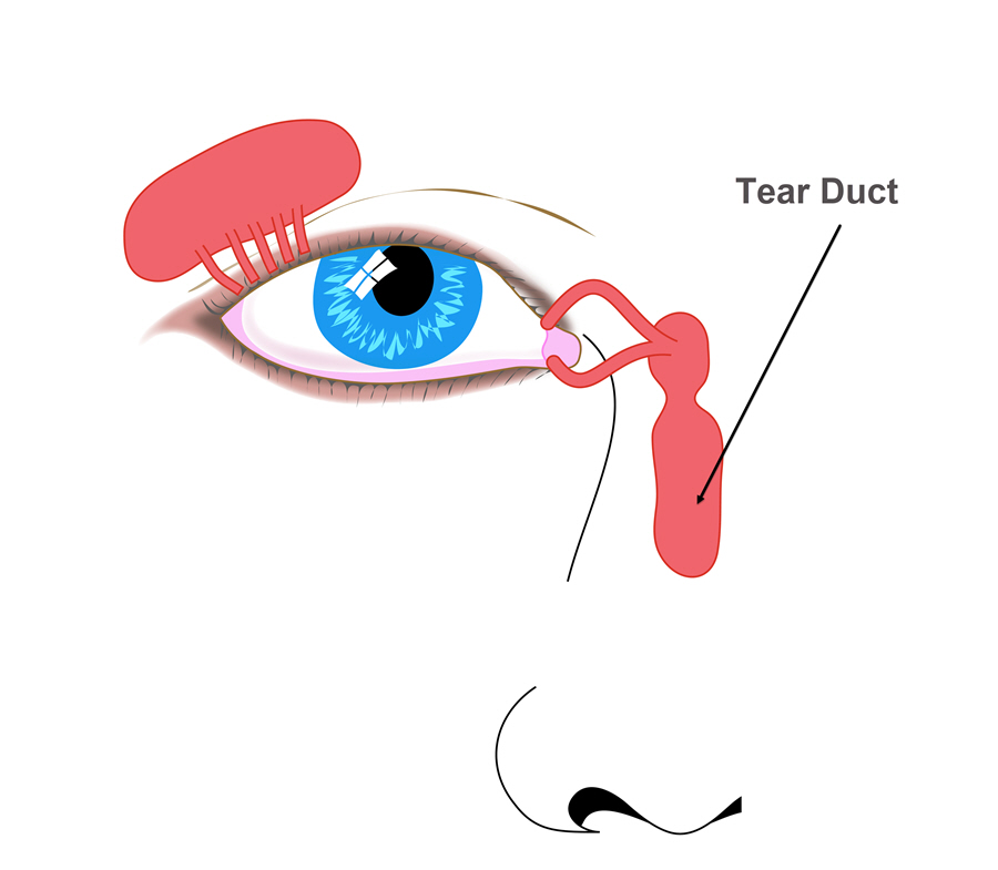
The tear duct is a continuous channel that connects to the tear (lacrimal) sac. It opens into the nasal cavity and forms the final part of the lacrimal apparatus. The lacrimal system (tear drainage system) is a set of linked anatomical structures situated within the eye orbit that handle the production and drainage of tears.
Also Known As
- Lacrimal duct
- Lachrymal duct
Anatomy
The tear duct has two parts:
- The intraosseous part – It is 12 mm long and enters the lacrimal groove and passes within the tear canal which is made by the lacrimal bone and the maxilla. It ends as a slit-like opening below the inferior nasal meatus. A mucosal fold known as plica lacrimalis (the valve of Hasner) covers the opening.
- The membranous layer – It is composed of connective tissue and stratified columnar epithelium.
Function
The purpose of the tear duct is to drains excess tears and waste material into the back of the nose through the nasal bone. When the eye blinks, the eyelid moves the tears and debris across the eye into the drains at the inner edge. The pipes flow into channels that link the eye with the nose. The channels empty into a tear sac which sits by the side of the nose. The tear sac links into the tear duct which drains into the nostril through the nasal bone.
Associated symptoms & disorders
Many problems can involve the tear duct. These include:
- Crusty eyelids and eyelashes – They occur when eye discharge dries on the lids and lashes. The condition may be associated with conjunctivitis (pink eye), blocked tear duct and blepharitis.
- Blocked Tear Duct – It happens when there is partial or total obstruction of tear drainage system. Tears accumulate in the eye causing excess tearing, irritation or chronic eye infection.
- Discharge from the eye – The eye discharge can be excessive tearing, mucus or pus. It could be linked with pink eye, blocked tear duct, corneal ulcer and keratitis among other eye diseases.
- Blurriness – It is a visual symptom which prevents a clear or sharp vision. Often, it is associated with many eye conditions including corneal disorders, retinal diseases, glaucoma, age-related macular degeneration (AMD), keratitis, etc.
- Tear duct infection (Dacryocystitis) – It occurs when there is a blockage of the tear drainage system. The blockage causes bacteria, fungi and viruses to collect in the lacrimal sac or the ducts.
- Swelling around eye – It is an inflammation that affects tissues around the eye and the eyelids. The disorder may be associated with many eye problems including black eye, blepharitis, eye allergies, blocked tear duct and conjunctivitis.
- Excess tearing – Increased tear production may cause excessive watering of the eyes. A blockage of the tear ducts close to the nose may result in excessive tearing from the eye. Excess tearing may also be linked with other condition such as Keratitis and corneal disorders.
Diagnosis of associated disorders
The eye care professional can diagnose tear duct disorders by reviewing the patient’s history and a comprehensive exam. The tests may include:
- Tear drainage test
- Slit lamp examination
- Irrigation and probing
- Fluorescein angiogram
- Ultrasound biomicroscopy
- Magnetic resonance imaging
- Optical coherence tomography
The professional also examines the patient’s nose to check for structural disorders that may be causing obstruction.
Treatment of associated disorders
The mode of treatment depends on the cause of the tear duct disorder. Often, infants and kids with the condition get better without treatment. The eye doctor may opt for careful observation. He/she may also use a particular massage technique that helps to open the valve of Hasner i.e. the tissue at the end of the duct. The doctor may prescribe medication to combat infection and nasal allergies.
Surgery may also be used in the treatment of tear duct conditions. The eye surgeon may use a wire to probe the tear duct and clear any blockage. It is a conventional technique for the treatment of recurring infections in infants.
The surgeon may use dacryocystorhinostomy, a laser procedure which expands a narrowed or blocked duct. He/she may have to remove the bone that causes the canal to narrow down.
Balloon catheter dilation is a technique that opens the tear ducts that are narrowed or blocked by scarring or inflammation.
In severe cases, the surgeon may have to reconstruct the whole tear drainage system.
