Introduction
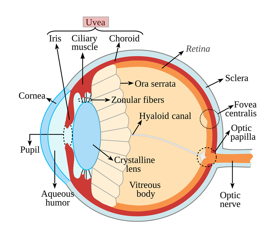
The uvea is the middle layer of the eye below the sclera. It is pigmented, consisting of the iris, ciliary body and choroid.
Also Known As
Vascular tunic
Anatomy
The iris is the colored part of the eye with the pupil lying in its middle. The iris and the pupil help control how much light enters the eye. The iris divides the anterior and posterior chambers of the eye. It is made up of connective tissue and muscle which surrounds the pupil. The iris determines the color of the eye due to the amount of pigment within it.
The ciliary body is located between the iris and the sclera (white part of the eye). It is invisible due to its location behind the sclera. It is also responsible for secreting the aqueous humor. This tissue measures about 6 or 7 millimeters wide in adults. It is divided into pars plicata and pars plana. Pars plicata has a thicker body while pars plana is flatter. The pars plana is attached to the choroid which lies on the posterior of the uvea.
The choroid sits between the sclera and the retina. It contains connective tissue and blood vessels.
Function
The uvea’s function is tied to the three components it is made of. These structures help adjust the eye to different levels of object distances and light.
The muscles in the iris cause the eye to constrict (narrow) in bright light and dilate (widen) in dim light. This action protects the eye from excessive light to enable vision in different environments.
The ciliary body keeps the lens in position through its attachment to the zonules. It also secretes aqueous humor and contains the muscle which controls accommodation.
The choroid nourishes the retina through its numerous, small blood vessels.
Associated symptoms & disorders
Some of the diseases that affect the uvea include:
- Uveitis which is inflammation of the uvea. Uveitis affecting the iris is called iritis while the one affecting the ciliary body is called iridocyclitis or anterior uveitis. Other types include intermediate uveitis, posterior uveitis and panuveitis (affecting all three areas of the uvea). It is not clear what causes the inflammation. Uveitis can lead to decreased visual acuity.
- Synechia is a disease in which parts of the iris form an adhesion at the front of the lens or the cornea’s back surface. It is caused by trauma to the eye, inflammation of the iris and so on. Synechia can lead to glaucoma.
- Iris coloboma occurs when a baby is born without a part of the iris. This gives the baby’s pupil a cat-like appearance. The disease can cause decreased visual acuity, blurred vision, double vision and ghost images.
- Uveal melanoma is cancer within the uvea. A choroidal melanoma occurs in the posterior uvea while iris melanoma occurs in the iris. The tumors develop in melanocytes, pigment cells located in the uvea. Uveal melanoma is the most common type of eye cancer. It can easily spread to other parts of the body.
- A choroidal nevus refers to a flat, benign pigmented area in the choroid. It is a freckle that the eye doctor needs to track to avoid complications.
- Choroideremia is an inherited, progressive degeneration of the choroid primarily affecting men. It can result in decreased visual field, night blindness and eventually blindness.
- Iris nevus is a freckle in the eye’s iris. In most cases, the freckle is harmless. However, an iris nevus can grow into cancer.
Diagnosis of associated disorders
The eye care professional diagnoses the various uvea-related conditions through the following:
- Visual field test
- Visual acuity test
- Fundus examination
- Complete medical history
- A thorough eye examination
- Ophthalmoscopy to examine the inside of the eye
- Blood tests and genetic testing for Choroideremia
- A slit-lamp exam with gonioscopy of angle structures especially for synechia
- Fluorescein angiography, fluorescein autofluorescence, electroretinography, optical coherence tomography and OCT angiography
Treatment of associated disorders
Scientists are yet to discover a cure for some of the conditions affecting the iris such as a coloboma. Instead, patients can wear prosthetic or colored contact lenses. These lenses give a round appearance to the eye. Surgery can correct the iris’ appearance.
Some conditions like a choroidal nevus and iris nevus require no treatment. The benign freckle should, however, be monitored in case it turns malignant.
Enucleation(removal of the eyeball) may aid in treating uveal melanoma.
Topical corticosteroids and dilating eye drops can assist with uveitis. The steroids treat the underlying cause. In some cases, eye injections and oral medications may be administered. Surgery to replace the vitreous or to implant a device for the slow release of steroid drugs is another option.
