Introduction
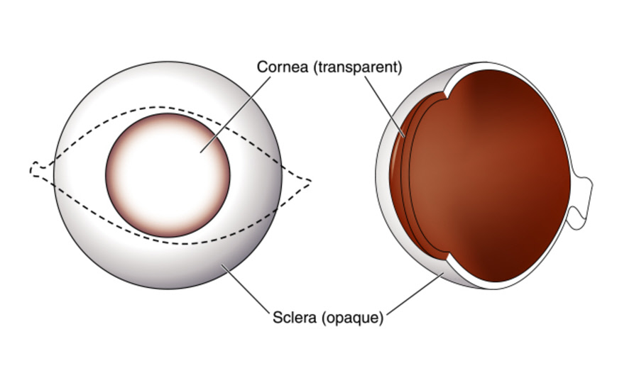
The sclera is the white part of the eyeball that extends back beyond the visible area of the eye. It is the thick outer cover that forms a tough coating for the eyeball. It encloses the front four-fifths of the eyeball surface. The sclera changes near the front the eyeball to become the cornea. It also forms the optic nerve dural sheath at the tail end of the eye.
Anatomy
The sclera has four layers:
- Episclera – A thin vascular tissue that lies between the sclera and the conjunctiva
- Stroma - A delicate mesh of fibers interspersed with blood vessels and nerves
- Lamina fusca - The layer of thin pigmented connective tissue on the inner surface
- Endothelium - The central layer of the endothelial cells
The conjunctiva covers the sclera. It is a clear mucus layer that aids in lubricating the eye. The conjunctiva is thickest around the optic nerve.
Function
The sclera protects the eyeball and provides structural integrity and rigidity. It is critical to the cohesion of the globe (eyeball of the eye without the external parts) and is resistant to most traumas. The sclera helps to maintain the shape of the eye and protects the vital eye components from injury. Furthermore, together with the retinal pigment epithelium and choroid, the sclera works to prevent unfocused light from reaching the retina.
Associated symptoms & disorders
The diseases that affect the sclera may be chronic, painful, collagen destructive and some may result in notable inflammation. Often, these conditions are localized indications of disease activities affecting the whole body. Disorders associated with the sclera include:
- Episcleritis – An inflammation of the episcleral membrane common in younger patients.
- Scleral Coloboma - A condition in which missing tissue can cause notching and bulging of the sclera.
- Scleral Ectasia – In this condition, the sclera becomes thin and bulges due to trauma, disease, atrophy or high intraocular pressure.
- Staphyloma - It is a condition in which the sclera develops a weak point and the internal eye pressure stretches it causing a protrusion.
- Scleritis – It is an inflammation involving the deep episclera and sclera. It is a severe, destructive and vision threatening condition.
- Blue sclera – It occurs because of the thinness and transparency of the sclera collagen fibers. It allows the veins in the underlying layer to show through.
- Melanosis - It is a genetic condition in which there are excess deposits of pigment (melanin) on the surface of the sclera. It can cause the sclera to become inflamed and uncomfortable.
Diagnosis of associated disorders
The eye care professional can diagnose scleral disorders by taking the patient’s medical records and eye tests. The tests may include:
- Blood test to check for signs of infection and immune system activity
- Slit-lamp examination - A routine procedure where the specialist shines a light into the eye to check for diseases or injuries
- Ultrasound Biomicroscopy (UBM) – It is a technique used for imaging of the front third of the eye
- Biopsy – A procedure which involves removing tissue from the sclera and examining it under a microscope
- Ultrasonography – It is a procedure which uses sound waves and allows the professional to look for changes occurring in or around the sclera
- Magnetic resonance imaging (MRI) – It is a non-invasive technique that gives 3D detailed anatomical images without using damaging radiation
Treatment of associated disorders
The method of treatment varies depending on the type of the disease. In genetic scleral disorders, the eye doctor may use symptomatic treatment to address the signs and symptoms. In other cases, the underlying causes determine sclera treatment. The doctor may prescribe medication such as:
- Antibiotics for infections of the sclera
- Systemic immunosuppressive therapy
- Oral glucocorticoids for posterior scleritis
- Antifungal medications for diseases caused by Sjogren’s syndrome
- Immunosuppressive drugs with oral glucocorticoids for necrotizing scleritis
- Nonsteroidal anti-inflammatory drugs in minor cases of nodular episcleritis
- Corticosteroid medication for eye pain, inflammation and vision protection
In severe cases, the eye surgeon may need to use surgery to repair damaged scleral tissues and prevent further loss of vision. Scleral transplant or grafting may be used where infection, erosion or other factors cause a weak area in the sclera and intraocular pressure creates a rupture. Scleral grafting may also be used to treat a deep penetrating wound caused by trauma.
