Introduction
Refractive surgery is a method of correcting or improving an individual’s vision. It is an option for people who no longer wish to wear glasses or contact lenses. Surgery helps to restructure the cornea (the transparent dome-shaped front surface of the eye). It also helps to adjust the focusing ability of the eye. Refractive surgery involves either reshaping the cornea or using surgical insertions.
Refractive surgery is for people with astigmatism, presbyopia (aging eye), hyperopia (farsightedness) and myopia (nearsightedness). However, people slated for refractive surgery should not be having any eye disease. They should also understand that they may need to use contacts and glasses after surgery to attain the best vision. Like any other surgical procedure, there are risks and potential side effects.
Types of Surgery
- Photorefractive keratectomy (PRK)
- Laser-assisted in situ keratomileusis (LASIK)
- Wave-front guided lasik
- Epi-LASIK
- Other refractive surgeries include:
- Lasek
- Conductive keratoplasty (CK)
- Phakic intraocular lenses (IOLs)
- Refractive lens exchange/Clear lens extraction/Clear lensectomy
- Small incision lenticule extraction (SMILE)
- Corneal inlays
- INTACS
Purpose
Refractive surgery, whether laser or non-laser, is aimed at reshaping the cornea. It improves the way light rays focus on the retina. Surgery corrects refractive errors to improve vision. Surgery also reduces or eliminates the need for contact lenses or eyeglasses.
Preparation and Expectation Before Surgery
Refractive surgery is ideal for those whose vision has stopped changing; generally for people above the age of 18. The following are the expectations before refractive surgery:
- The patient’s vision must not have changed in that particular year
- The cornea is healthy
- Not having corneal abrasions, diabetes, advanced glaucoma, cataract and eye infections
- Not having a history of scarring
- Not being pregnant or nursing
- Having realistic expectations about the outcome of surgery
Generally, the eye doctor will conduct the following procedures prior to surgery:
- Test vision - To check that vision has not changed and ascertain the exact refractive error
- Check for eye problems - Surgery cannot be done with existing eye problems. Surgery could worsen these problems or the problems may interfere with the operation.
- Measure the thickness and surface of the cornea using topography, tomography and pachymetry tests. He/she then enters this data to a computer-based laser.
Measure the size of the pupil and check the optic nerve
Procedure
1. Photorefractive Keratectomy (PRK)
PRK is an outpatient procedure that lasts about 15 minutes. The surgeon will numb the eyes with eye drops. He/she will keep the eye from blinking by using an eyelid holder. The ophthalmologist will then remove the epithelium (outer layer of the cornea's cells). He/she removes the epithelium using an alcohol solution, a blade, a special brush or laser. The ophthalmologist will request the patient to look intently at a target light to avoid eye movement. The cornea will then be reshaped using a laser and a ‘bandage' contact lens will be placed over the eye.
2. LASIK
Wavefront-Guided LASIK
In Wavefront-Guided LASIK, a computer imaging technology designs an exhaustive three-dimensional map of the cornea. The excimer laser is then programmed with the patient’s wavefront data. The technology ‘sculpts’ the cornea to correct vision.
Epi-LASIK
The surgeon will use the Epi-keratome to accurately separate an extremely thin leaf of epithelial tissue from the cornea. He/she will shift the sheet to the side. The surgeon will then conduct PRK. He/she has the option of re-attaching the thin sheet to the cornea or removing it. Afterwards, the surgeon applies a ‘bandage’ soft contact lens.
3. Other Surgical Procedures
Lasek
A trephine (microsurgical instrument) creates a corneal tissue epithelial flap. The professional will use an alcohol solution to relax the epithelial cells. At this point, a PRK procedure will be performed. After sculpting the cornea, he/she will reposition the epithelial flap. He/she will then smooth the epithelial flap with a tiny spatula and secure it with a ‘bandage’ of soft contact lens.
Conductive Keratoplasty (CK)
The professional will use a small probe that gives out controlled amounts of radio frequency energy in this non-invasive thermal procedure. This procedure does not use laser techniques. CK applies heat to the marginal part of the cornea causing it to reduce in size and tighten. The shrinking and tautening will increase the cornea’s curvature and improve the optical power of the central cornea.
Phakic Intraocular Lenses (IOLs)
In Phakic IOLs the implant contact lens will be surgically implanted. They are placed inside the eye in front of the natural lens. The exact location is in front of or behind the iris. The ophthalmologist does not remove the natural lens of the eye.
Refractive Lens Exchange/Clear Lens Extraction/Clear Lensectomy
The eye’s natural lens are replaced with an artificial lens. It is a procedure similar to cataract surgery. He/she may use accommodative intraocular or multifocal lenses to allow focusing at all distances. Early stage cataract patients and people with hyperopia can opt for it.
Small incision lenticule extraction (SMILE)
The ophthalmologist will use a laser to cut a small lenticule (disk) of corneal tissue. This tissue is then removed through a tiny incision (2 to 4 mm) in the adjacent cornea.
Corneal Inlays
To treat presbyopia, corneal inlays (implants) are used. The implants are placed into the center of the cornea in front of the pupil. The implant is put under a flap of tissue made by a laser or a microkeratome (a cutting device).
INTACS
Small plastic arc-shaped segments are implanted into the middle layer of the cornea near the outer edge.
After Care, Recovery, Results
The patient may experience blurry vision after laser treatment. The healing period depends on the condition and treatment. For example, patients who have had a PRK will have their vision improve gradually over 3-5 days. It may take a month or longer to attain the best vision. 90% of patients who undergo refractive surgery improve to 20/40 vision or better without having to use contacts or glasses. However, people over 40 years old will need reading glasses even if they don’t have to wear eyeglasses after refractive surgery.
Wavefront LASIK may reduce side effects such as night vision. Those who go through Lasek and Epi LASIK take about four days for healing of the epithelial layer to take place. CK and PRK can assist presbyopia patients to achieve monovision (one eye is allowed to stay at nearsightedness while the other one is adjusted to see distant objects).
The eye doctor will require the patient to do the following:
- Have someone drive them home soon after surgery
- Rest, take a nap and relax
- Take a few days from work
- Avoid strenuous activities for a week
- Use over-the-counter medicine to help with pain
- Use eye drop medication for up to a month or as directed by the doctor
- Wear sunglasses according to the doctor’s instructions
Risks & Complications
The risks or complications of refractive surgery include:
- Infection of the cornea
- Scarring of the cornea
- Sensitivity to light especially at night
- Glare and halos around lights
- Corneal haze (cloudy cornea)
- Under correction or overcorrection of vision
- Wrinkling of the cornea or deposition of cells inside the cornea
- Very rarely, a patient may experience worse vision
- Very infrequently, blindness
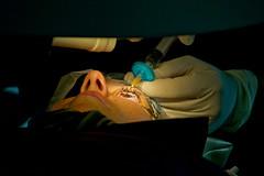
Image by Peretz Partensky from San Francisco, USA - Lasik : Laser Eye Surgery, CC BY 2.0, https://commons.wikimedia.org/w/index.php?curid=24351064
