Introduction
The retina is the thin light-sensitive nerve membrane lining the interior of the eyeball. It lies near the optic nerve. The retina and the optic nerve are outgrowths of the developing brain. Thus, they are brain tissue and are considered components of the central nervous system.
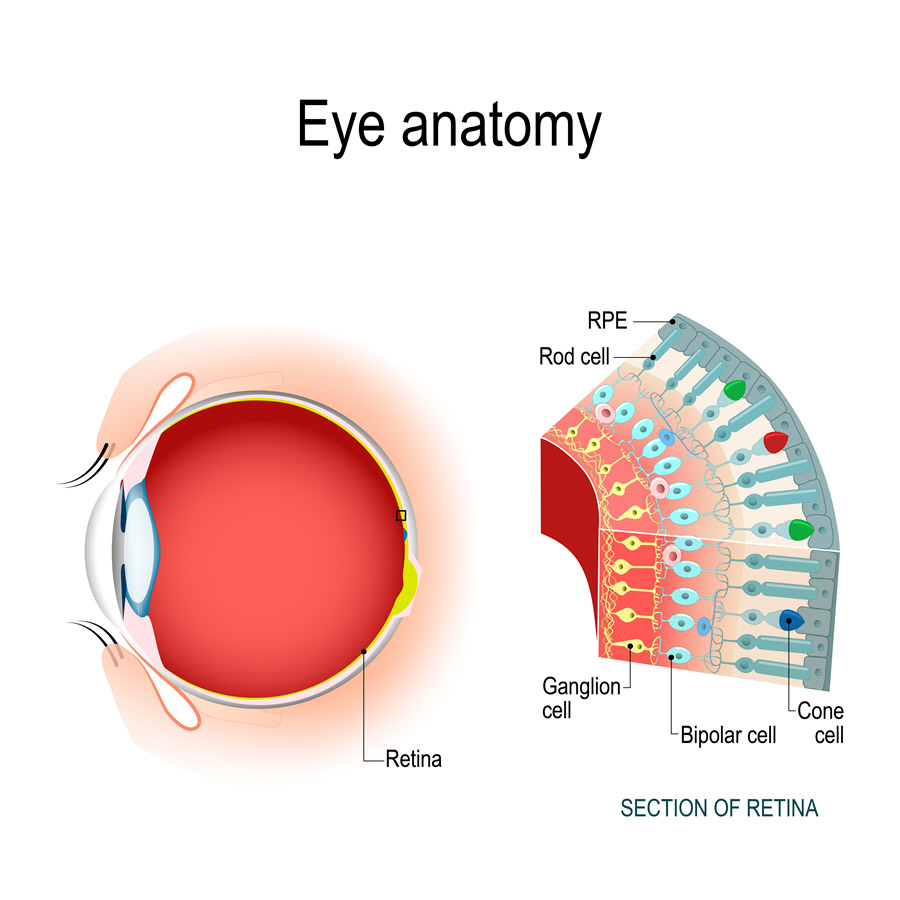
Anatomy
The retina has three main layers:
- Photoreceptors made of rods and cones
- Bipolar cells
- Ganglion cells
These parts are connected through intermediate tissues of:
- Horizontal cells
- Amacrine cells
The macula and the fovea lie at the center of the retina. The macula provides the central vision needed for fine details, reading and driving. The fovea is composed of the cone cell which gives the sharpest image. The retina receives oxygenated blood (nourishment) from the choroid.
Function
When light enters the eyes, the eye lens focuses it onto the retina. The retina converts it into neural impulses and transmits the messages to the brain for visual interpretation. In other words, the retina processes an impression from the lens, and the brain decides what it is.
The retina uses the layer of photoreceptor cells to process the incoming light. These are special light-sensitive cells that detect exceptional image qualities such as color and light intensity.
Associated symptoms & disorders
Many problems can affect the retina and lead to loss of vision. Some of these conditions include:
- Macular pucker – A scar tissue forms on the macula
- Retinitis pigmentosa – It is a retinal degenerative disease
- Macular hole – It is a small defect in the middle of the retina
- Macular degeneration – The center of the retina begins to deteriorate
- Retinoblastoma – It is a form of cancer that develops from the immature cells of a retina
- Cytomegalovirus retinitis – It is an inflammation of the retina that can lead to blindness
- Central serous retinopathy – It is a disorder where the retina detaches and causes vision loss
- Choroidal neovascular membranes – In this condition, new damaging blood vessels begin to grow beneath the retina
- Epiretinal membrane – The epiretinal membrane which lies on top of the retina can pull it up and cause distorted vision
- Retinal detachment – The retina separates from the underlying layer of tissue due to an accumulation of fluid under the retina
- Macular edema – It is the buildup of fluid in the macula. It occurs due to leakage from damaged blood vessels in the retina
- Retinal tear – The retina breaks as a result of traction because of the shrinkage of the vitreous. The vitreous is the clear, viscous material occupying the center of the eye.
- Diabetic retinopathy – It affects people with diabetes. In this condition, the blood vessels at the rear of the eyeball deteriorate causing fluid to leak under and into the retina. It causes the retina to swell and can blur or distort the patient’s vision.
Diagnosis of associated disorders
The eye care professional can diagnose retinal disorders by reviewing the patient’s medical record and a comprehensive eye exam. The tests may include:
- Blood tests
- Tonometry
- Color testing
- Dilated eye test
- Visual field testing
- Retinal photography
- Fluorescein angiography (FA)
- ERG (Electroretinography) test
- Optical coherence tomography (OCT)
- Magnetic resonance angiography (MRA scan)
- Computed tomography angiography (CTA scan)
Treatment of associated disorders
The form and extent of the retinal disorder determine the method of treatment. The treatment may involve medication and surgery.
Medication may stop or even reverse the effects of inflammation and the development of abnormal blood vessels. The eye doctor may prescribe anti-vascular endothelial growth factor (anti-VEGF) therapy or steroid treatment. In anti-VEGF treatment, the doctor administers injections with antibodies to block the VEGF. It helps to stop the leakage and bleeding of fluids from the damaged retinal blood vessels. Steroid treatment may be used where the anti-VEGF therapy is not viable, for example, in patients who have had cataract surgery.
In severe cases, the eye surgeon may use laser therapy to stop the leakage of fluid and blood in the retina. He/she uses a laser to seal the leaking blood vessels and stop more fluid from entering the retina. The surgeon may also use vitrectomy to repair detached retinas and to remove fluid from inside the eye and scar tissue on the retina.
