Introduction
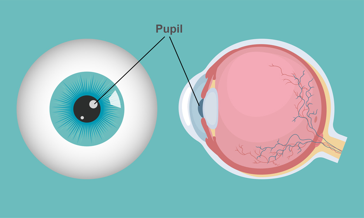
The pupil is the opening in the middle of the iris. Light passes through the pupil before reaching the lens. The size of the pupil is adjusted by the iris which also controls how much light enters the eye.
Anatomy
From the cornea, light passes through the pupil before it is focused on the retina. The muscles in the iris control the movements of the pupil. With bright light, the pupil constricts (narrows) and dilates (widens) in dim light.
Parasympathetic nerve fibers affect the muscle that causes the pupil’s constriction while sympathetic nerve fibers control dilation. The pupil contains the pupillary aperture which narrows with close objects (accommodative pupillary response) and widens in objects at a distance.
Pupil sizes vary from small to large. Age, disease, trauma and other abnormalities may affect the size of the pupil.
An average pupil measures 2 to 4 millimeters in diameter (in bright light) and 4 to 8 millimeters (in dim light). The pupils are round and have the same black color and size. The light that passes through the pupil is not reflected hence the black color.
Function
The pupil allows light into the eye so that it can be focused onto the retina and ultimately provide vision. Together with the iris, it controls the amount of light entering the eye.
Associated symptoms & disorders
Some of the eye problems that affect the pupil include:
Anisocoria where the patient has unequal pupil size. The disease may be congenital or caused by nervous system disorders. With anisocoria, focusing on near objects becomes a challenge.
Pupil-white spots refers to pupils which have white spots giving the pupil a white instead of black color. The condition can cause decreased vision even before the pupil shows the white color.
Coloboma of the iris is a defect of the iris, often congenital. It may appear as a second pupil giving the pupil an irregular shape. It causes blurred, decreased and double vision.
Adie's tonic pupil is a condition in which the pupil shows almost no reaction to light. There is also a delayed reaction to accommodation (the eye’s ability to change its focus from distant to near objects and vice versa). The disease involves typically one eye where the concerned pupil is larger than the healthier iris.
The rare Argyll Robertson pupil involves an unresponsive pupil to light but whose reaction to accommodation is normal. It affects both eyes with both pupils being too small to react to light. The cause remains unknown but it has been associated with diabetic retinopathy and syphilis.
Trauma can affect the pupil especially penetrating eye trauma. It is responsible for pupils that have an abnormal shape. Injury can also be a consequence of cataract surgery complications, refractive lens exchange or phakic intraocular lens (IOL) surgery. The pupillary response to light is affected.
Horner’s syndrome is a condition where one pupil is smaller and fails to dilate as it should. It can lead to ptosis (drooping of the eye) of the upper eyelid.
Diagnosis of associated disorders
The eye care professional may do the following to diagnose the diseases associated with the pupil:
- Ophthalmoscopy which examines the back of the eye
- Do a physical examination which includes a detailed eye examination
- Visual acuity test to see how far one can read from a chart a distance away
- A slit-lamp exam to examine the structures located at the front of the eye
- Take medical history especially information on smoking in the case of anisocoria
- Imaging tests such as computed tomography and magnetic resonance imaging
- A standard eye examination which includes checking how the pupil responds to light
Treatment of associated disorders
Some of the diseases such as anisocoria and Horner syndrome may require no treatment. Only the underlying causes need to be treated.
There is no cure for a coloboma. Patients affected by iris coloboma may put on colored contact lenses. These lenses make the iris look round.
For Adie's tonic pupil, reading glasses can assist with vision. The patient applies Pilocarpine drops thrice a day to help with the constriction of the dilated pupil. The treatment aids in depth perception and reduction of glare.
There is no treatment for Argyll Robertson pupil. However, the progression of the disease can be controlled when the underlying disease, syphilis, is treated. Treatment usually involves penicillin G benzathine.
Surgery can also correct the appearance of the iris.
