Introduction
Photoreceptors are specialized cells located in the retina, the light-sensitive tissue at the back part of the eye. Rods and cones make up the two types of photoreceptor cells.
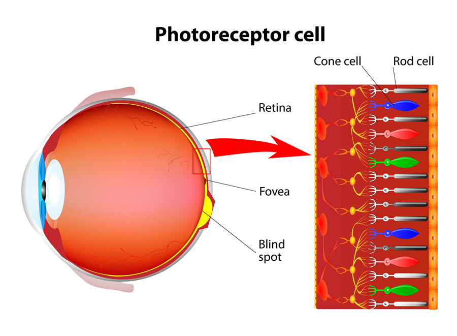
Anatomy
The retina is made up of the central retina and peripheral retina. The fovea and the macula occupy the central retina. The fovea (the center of the macula) provides clear, sharp vision. It only contains cones, packed tightly together than in any other area of the retina. Compared to rods, cones are less sensitive to light.
About 6 million cones inhabit the human retina. Cone cells come in three types: red-sensing cones (60%), green-sensing cones (30%) and blue-sensing cones (10%). When the brain receives signals from the brain, it translates them into color perception. However, some people cannot perceive some colors because of the absence of a particular cone. It could also be that one type of cone is weak. Such individuals are said to be color blind. Cones also help to detect better, detailed vision. They work best in bright light.
The retina contains about 120 million rods. They provide peripheral vision since they are numerous on the side of the retina. Rods have more sensitivity to light than cones. They are also sensitive to movement and shape. Rods enable one to see in a dim room.
Function
Photoreceptors perform the unique role of converting light into signals which are then sent to the brain. They provide color, night and peripheral vision.
Associated symptoms & disorders
Several eye problems can affect cones some of which include:
- Color blindness (color deficiency) is a congenital disorder when an individual fails to perceive colors in both eyes the normal way. The individual is unable to distinguish between certain colors, often red and blue. The absence of color cones, a malfunction of the color cone or a cone that detects a different color than usual can lead to color blindness. People with mild color blindness have all three cone cells except that one has malfunctioned. Those with severe color deficiencies have all the three cones missing. A rare form of severe color blindness occurs when everything is seen in shades of gray.
- Photokeratitis occurs when the eye is exposed to ultraviolet (UV) rays. UV rays can come from the sun or man-made sources. Photokeratitis affects the cornea, conjunctiva and the eyelids. The disease can lead to temporary color changes in one’s vision.
- Retinitis pigmentosa is a group of eye problems associated with the retina. It alters how the retina responds to light giving rise to decreased vision. One gradually loses vision though the disease does not cause blindness. Some people may also have trouble seeing certain colors.
- Usher syndrome is a genetic autosomal recessive disorder. The disease causes loss of hearing and an eye disorder called retinitis pigmentosa (RP). RP is a group of eye problems affecting the retina. RP alters how the retina responds to light leading to difficulties in vision. Usher syndrome can result in loss of color perception when the rods begin to degenerate.
- Other diseases affecting photoreceptors may include rod-cone dystrophy, Stargardt disease, etc.
Diagnosis of associated disorders
For color blindness, the eye care professional requires one to read from the Ishihara color plates. It is a pattern composed of multi-colored dots and numbers written inside it. Those without color blindness will spot the numbers and shapes among the dots. Colorblind patients may fail to see anything in the pattern.
To diagnose photokeratitis, the professional will inquire about an individual’s recent activities. The eye is examined and an eye drop and fluorescein dye are used to check if UV rays have damaged the eye.
Retinitis pigmentosa and Usher syndrome are diagnosed through genetic testing, electroretinography, visual field testing and optical coherence tomography (OCT). Usher syndrome can further be diagnosed through an audiology test.
Treatment of associated disorders
Researchers are still trying to find treatment for congenital color blindness. Special contact lenses and glasses can aid in providing the missing colors. The disease does not pose any threat to vision. Doctors treat acquired forms of color blindness by addressing the underlying cause such as retinal and optic nerve disorders.
Photokeratitis usually goes away on its own. Treatment is focused on making one better as the eye heals. The patient can use a cold washcloth, artificial tears, painkillers and eye drop antibiotics.
Doctors are yet to find a treatment for retinitis pigmentosa. Currently, patients may take vitamin A, including vitamin A palmitate to slow the progression of RP.
Scientists are still investigating a cure for Usher syndrome. Treatment involves giving the patient valuable education to enable them to maximize on both hearing and vision aids. Patients receive braille and sign language instruction. They also get mobility training (low-vision devices and techniques).
