Introduction
The eye is divided into various parts. Each section plays a significant role in enabling vision. The components of the eye work together to provide vision. A malfunction or disease in any part of the eye can affect vision.
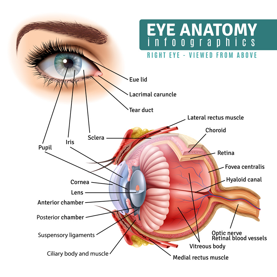
Anatomy
- The following are some of the major parts of the eye:
- Orbit
It is the socket made of bones whose job is to protect the eye. The eye is located inside the orbit.
- Muscles
There are six extraocular muscles which aid with eye movement. They move the eyes up, down, side to side and help rotate the eye.
- Sclera
It’s a strong layer of tissue which covers the entire surface area of the eye. The sclera is the white part attached to the extraocular muscles.
- Eyelid
The eyelid is folded skin that closes over the eye. Its function is to protect the eye. It is composed of the upper and lower eyelid.
- Conjunctiva
It is a transparent membrane which covers the surface of the eye and the eyelid’s inner surface.
- Anterior chamber
It is the part that is filled with aqueous humor and located behind the cornea. The aqueous humor drains from the eye through the drainage angle, maintaining eye pressure.
- Cornea
It is the transparent membrane on the outer part of the eye. Light initially falls on the cornea before it is sent to the retina.
- Iris
It is the colored part of the eye. The pupil occupies the center of the iris. The iris regulate show much light enters the eye.
- Lens
It is a transparent area of the eye and positioned at the back of the iris. It assists in focusing light and images on the retina.
- Retina
It is a light-sensitive tissue that occupies the back of the eye. The retina receives focused light, converts it into neurons and sends it to the brain via the optic nerve. The photoreceptor cells in the retina alter light to energy which is then sent to the brain. Photoreceptor cells are divided into two; rods and cones. Rods discern black and white and provide night vision while cones discern color and give detailed vision.
- Macula
The macula occupies the center of the retina. It is responsible for the central vision in the eye. The other part of the retina is called peripheral retina which provides the eye’s side vision.
- Optic nerve
It is the largest sensory nerve of the eye. Its task is to carry sight impulses from the retina to the brain.
- Pupil
Light passes through the pupil, located in the middle of the iris. The iris controls the amount of light entering the eye by narrowing (in bright light) and widening (in dark or dim light) itself.
- Vitreous humor (gel)
Two-thirds of the rear part of the eyeball is filled with vitreous gel. Vitreous humor is a clear, colorless mass located between the lens and the retina. It nourishes the inside of the eye and helps in shaping the eye.
Function
The human eye functions to provide vision. Various parts of the eye such as the cornea, lens, retina and so on need to be healthy to provide vision.
Associated symptoms & disorders
Each of the parts can malfunction or be affected by certain conditions leading to eye diseases. The following are some of them:
- Corneal-related diseases can be caused by injuries, allergies, dry eye and inflammation of the cornea (keratitis). These can lead to corneal scars or abrasions which threaten vision. Corneal dystrophies are usually inherited and can cause little to severe vision impairment. They include keratoconus, Fuch’s dystrophy, lattice dystrophy and map-dot finger dystrophy. Other diseases that can affect the cornea include shingles (herpes zoster), ocular herpes, pterygium, iridocorneal endothelial syndrome and Stevens-Johnson syndrome.
- The most common conjunctival and scleral disorders are inflammatory and include conjunctivitis, scleritis and episcleritis. Conjunctiva-related diseases can be acute or chronic and result from infections, allergies or irritants. Scleritis and episcleritis are immune-related diseases.
- Retinal diseases affect any part of the retina and can lead to loss of vision. They include a retinal tear, retinal detachment, diabetic retinopathy, epiretinal membrane, macular hole, macular degeneration, retinitis pigmentosa, etc.
- Diseases associated with the iris include iritis, ocular melanoma, aniridia, coloboma, plateau iris, etc. These conditions can cause distorted, blurry, double vision and blindness.
- The most popular disease related to the lens is a cataract. It occurs when the lens becomes cloudy or opaque.
- Muscle eye problems can emanate from weak muscles or a problem with the nerve that controls the muscle. They include diplopia, myasthenia gravis and graves’ disease. They can cause double vision and drooping eyelids.
- Congenital or hereditary defects affect the eye too such as coloboma and albinism.
- Cancerous (retinoblastoma, ocular melanoma) and non-cancerous (hemangioma) tumors can affect the eye.
- Refractive diseases include nearsightedness, farsightedness and astigmatism.
Diagnosis of associated disorders
Diagnosis depends on the condition. Diagnostic test options may include a visual acuity test, color test, field vision test, imaging tests, slit-lamp examination, blood tests, etc.
Treatment of associated disorders
Some conditions require no treatment and will resolve on their own such as viral conjunctivitis. Doctors are yet to find a cure for diseases like coloboma. Instead, patients are taught to manage the condition using such treatments as low-vision devices and so on.
Refractive errors are corrected using eyeglasses, contact lenses and laser surgery.
Medical treatment and other symptom-relieving medications may include topical steroid eye drops, dilating eye drops, anti-inflammatory drugs, painkillers, artificial tears, etc.
Surgical options include cataract surgery, corneal transplantation and enucleation (removal of the eyeball).
Laser surgery includes LASIK, thermal laser surgery, cryopexy, etc.
