Introduction
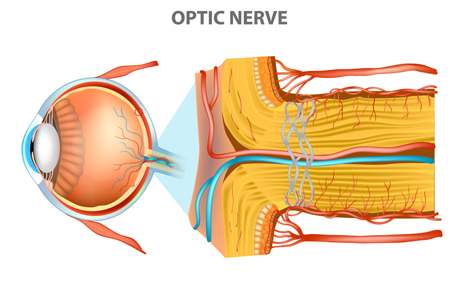
An optic nerve is a bundle of optic fibers that travel from the eye to the brain. The fibers are composed of glial cells and axons of retinal ganglion cell. They converge at the optic disc at the posterior pole of the eyeball and form the nerve. The optic nerve then moves towards the optic chiasm where fibers splits into optic tracts as others cross. Each eye has a single optic nerve that carries about a million fibers.
The optic nerve is the second cranial nerve in the peripheral nerve system. It is considered part of the central nervous system because:
- The optic fibers are myelinated by the oligodendrocytes. The fibers of the peripheral nerves are myelinated by the Schwann cells.
- They are encased within meningeal layers
- They are also not regenerated after damage as is the case with peripheral nerves
Also Known As
- Nervus opticus
- Cranial nerve II (CN II)
Anatomy
The optic nerve runs about 4 cm in length. It extends from the eyeball to the middle cranial fossa of the brain. It can be divided into four distinct parts:
- Intraocular part– The optic nerve begins at the optic disc/nerve head located at the eyeball. The retinal neural fibers converge at this point, passing through the surface nerve fiber layer, the choroidal/glial region and the lamina cribrosa. The fibers come out myelinated by the oligodendrocytes. They also get encircled by the meninges in the extraocular space
- Intraorbital part – The bundle goes to the orbital space, that is the eye socket after leaving the optic disc. The diameter of the intraorbital part is twice that of the intraocular due to the sheath covering. Here, the nerve is surrounded by three meningeal layers; the arachnoid, pia mater and dura
- Intracanalicular part – This is the part within the optic canal of the sphenoid bone. It allows additional movement of the eyeball within the orbital space.
- Intracranial part – This portion extends from the optic canal up to the middle cranial fossa where the two optic nerves from both eyes unite. The nerves create the optic chiasma. From there, the nerves diverge as optic tracts.
Function
The optic nerve runs about 4 cm in length. It extends from the eyeball to the middle cranial fossa of the brain. It can be divided into four distinct parts:
Intraocular part– The optic nerve begins at the optic disc/nerve head located at the eyeball. The retinal neural fibers converge at this point, passing through the surface nerve fiber layer, the choroidal/glial region and the lamina cribrosa. The fibers come out myelinated by the oligodendrocytes. They also get encircled by the meninges in the extraocular space
Intraorbital part – The bundle goes to the orbital space, that is the eye socket after leaving the optic disc. The diameter of the intraorbital part is twice that of the intraocular due to the sheath covering. Here, the nerve is surrounded by three meningeal layers; the arachnoid, pia mater and dura
Intracanalicular part – This is the part within the optic canal of the sphenoid bone. It allows additional movement of the eyeball within the orbital space.
Intracranial part – This portion extends from the optic canal up to the middle cranial fossa where the two optic nerves from both eyes unite. The nerves create the optic chiasma. From there, the nerves diverge as optic tracts.
The optic nerve transmits visual information from the eyes to the brain for interpretation. As the fibers split at the optic chiasma they proceed to different nuclei as follows:
- Lateral geniculate nuclei – The visual afferent fibers travel to the lateral geniculate body. They are responsible for transmitting visual impulses, enabling perception of images.
- Pretectum nuclei – The pupillary afferent fibers move to this body. They are tasked with pupillary light reflex.
- Superior colliculus nuclei – The photostatic fibers move toward the superior colliculus of the midbrain. They control movement of the eyes.
- Suprachiasmatic nuclei – Other fibers travel to this body. The fibers control hormonal changes and diurnal rhythms.
Associated symptoms & disorders
The main disorders that affect the optic nerve are:
- Glaucoma – The disease is associated with a build up of internal eye pressure. The elevated pressure can cause damage to the optic nerves if not controlled. The condition begins with loss of peripheral vision due to the loss of retinal cells. It then proceeds to the optic nerve.
- Anterior ischemic optic neuropathy (AION) - This is another common disorder of the optic nerve. It involves damage to the optic disc due to disruption of blood flow. Loss of vision can occur suddenly upon waking up.
- Papilledema – This is a condition of swelling of the optic disc. It may be bilateral or unilateral. Bilateral papilledema is known to occur due to intracranial pressure. The unilateral form could arise from primary tumor of the optic nerve.
- Optic nerve hypoplasia – This is the underdevelopment of the optic nerve. It may result in diminished to no vision in the affected eye
- Optic neuritis – Inflammation of the optic nerve could occur due to injuries, infections or underlying diseases such as multiple sclerosis.
Diagnosis of associated disorders
Diagnosis is necessary to evaluate the health of the optic nerves. Signs that could indicate a condition of the optic nerves are:
- Edema/swelling of the nerve
- Hemorrhage
- High intraocular pressure
- Loss of side vision
- Complete vision loss
- The most common tests performed to assess the condition of the optic nerves include:
- Ophthalmoscopy test – Involves the use of an ophthalmoscope to examine the inside of the eye.
- Biomicroscopy – A slit lamp and a special lens are used in this test to have a view of the posterior portion of the eye
- Visual field and acuity tests - The field test assesses the degree of visual field. It can detect loss of peripheral vision. An acuity test evaluates the clarity in vision.
- Tonometry – Where glaucoma or ocular hypertension is suspected, a tonometry test can be taken to check the pressure level. Pressure above 21 mmHg is considered high and can result in optic nerve atrophy.
Treatment of associated disorders
Majority of the conditions require proper monitoring by eye care doctors. Medications that are prescribed may include:
- Corticosteroids – They are issued where there is inflammation such as in optic neuritis or in the arteric form of anterior ischemic optic neuropathy.
- Glaucoma drugs – These include beta-blockers, carbonic anhydrase inhibitors and alpha-adrenergic agonists. They work to reduce intraocular pressure by either inhibiting fluid production or improving drainage.
- Other forms of medication such as pain killers can be issued where there is pain. Some conditions affecting the optic nerve require treatment of the underlying condition. Where infections are involved, antibiotics or antivirals may be prescribed. Surgery could also be indicated when a tumor is detected.
