Introduction
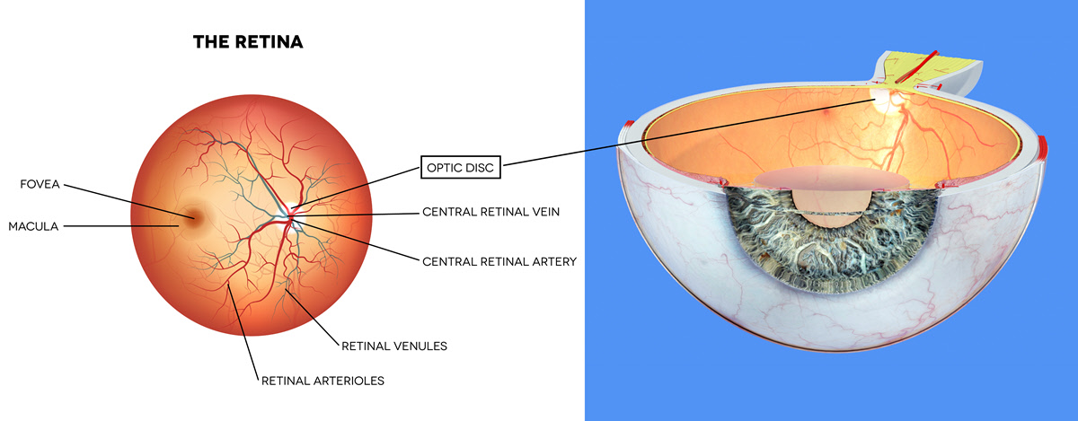
The optic disc is the part of the human eye where the axons of the ganglion cells converge and exit the retina. It contains no photoreceptors and therefore does not respond to light stimuli. It represents the blind spot in the eye.
About 1.2 million retinal nerve fibers come together at the optic disc. This is also the starting point of the optic nerve, hence the name optic nerve head.
Also Known As
- Optic nerve head
- Blind spot
- Discus nervi optici
Anatomy
The optic disc is a vertical-oval tissue that measures approximately 1.5mm in diameter. It contains a slight depression at the center referred to as the optic cup. The cup lies horizontally within the disc. In a healthy eye, the cup to disc ratio is less or equal to 0.7.
The optic nerve head has four distinguishable parts which are:
- The surface nerve fiber layer – This is the most anterior part of the head. It is continuous with the nerve fiber of the retina.
- Glial prelaminar region – The prelaminar region comes after the surface nerve fiber layer. It consists of bundles of nerve fiber surrounded by sheath.
- Laminar area – This is the scleral portion which contains the Lamina Cribrosa. It is made up of tissues with perforations for passage of the nerve fiber bundles and retinal blood vessels.
Retrolaminar portion – This is the posterior part of the optic disc. It shares blood supply with the optic nerve.
Function
The optic disc is the entry point for not just the optic nerve but also the retinal blood vessels. The optic nerve transmits visual signals from the eye to the brain. The brain then interprets the information and converts it to the images seen.
The retinal blood vessels, on the other hand, are responsible for supplying the retina with the oxygen and nutrients needed. The central retinal artery and vein pass through the optic cup to get to the retina.
Associated symptoms & disorders
The disorders/diseases associated with the optic disc include:
- Papilledema – This is the swelling of the optic nerve head. The edema is caused by a buildup of intracranial pressure, that is, pressure in/around the brain. The pressure may rise after a head injury, bleeding in the brain or infections. Symptoms of the condition can include blurry vision, headache and vomiting.
- Optic disc drusen - It can also be referred to as pseudopapilledema due to how the condition simulates features of edema. Optic disc drusen is a disorder where there’s calcification of globules of protein-like material and sugar molecules in the optic disc. The deposits cause the nerve head to elevate. The condition can result in loss of visual field and acuity.
- Optic neuritis – This is a condition of inflammation of the optic nerve. It can occur due to trauma, injury or infection.
- Anterior ischemic optic neuropathy – Ischemic optic neuropathy is damage to the optic nerve due to poor blood supply. When the optic nerve head is affected, it is referred to as anterior ischemic optic neuropathy.
- Glaucoma – Most glaucoma cases arise from high intraocular pressure. The pressure, if not controlled, can cause damage to the optic nerve.
Diagnosis of associated disorders
Diagnosis for diseases of the optic disc can include tests such as:
- Ophthalmoscope – This is used to view the interior of the eye. Light is shone inside to make it possible to see.
- Dilation exam – A dilator drug is placed on the eye to widen the pupil.
- Biomicroscopic exam – It involves use of a slit lamp together with a special type of lens to view the back of the eye. In most instances, dilator drops are used to make the diagnosis more accurate.
- Imaging tests – They include a fundoscopy, optical coherence tomography (OCT), magnetic resonance imaging (MRI) and retinal imaging.
The diagnosis can show the following signs of disease:
- Swollen optic disc – A healthy eye disc is flat. Any form of swelling can indicate disorders such as anterior ischemic optic neuropathy or papilledema.
- Hemorrhage – Glaucoma can show hemorrhages in some cases.
- Optic nerve cupping – This is where the cup to disc ratio is greater than normal. When the ratio exceeds 0.7, there is usually an underlying problem such as glaucoma.
- Blurred disc contours – In conditions such as optic disc drusen, the edges of the disc are not well defined.
- Afferent pupillary defect – When light is shone to one eye at a time, the pupils may respond differently, what is called afferent pupillary defect. It happens when the disease of the optic nerve is asymmetrical/unilateral. The diseases can include optic neuritis or anterior ischemic optic neuropathy.
Treatment of associated disorders
Not all disorders of the optic disc require treatment. Some like optic disc drusen will easily resolve on their own. Non-arteritic ischemic optic neuropathy is also not treated with conventional medication. Some of the treatments used in optic nerve head disorders are:
- Beta-blockers - They are used in glaucoma cases to reduce the intraocular pressure.
- Corticosteroids – In cases where there’s inflammation such as in optic neuritis, corticosteroids can be prescribed.
- Pain-killers – They are issued in cases of severe pain.
In papilledema where the cause is idiopathic intraocular hypertension, the patient may be required to start on a weight loss plan or a low-salt diet. In other instances, where the underlying problem could be a brain tumor, infection, bleeding or a clot, surgery may be required.
