Introduction
The lens is the clear part of the eye that is located behind the iris. Its sole purpose is to focus the light entering the eye onto the retina. The retina will then transmit those images to the brain resulting in vision. The lens enables the eye to focus on objects that are both close and distant.
Also Known As
Crystalline lens
Anatomy
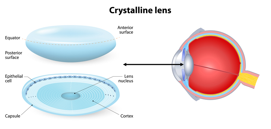
The lens has a sphere-like, biconvex shape. It measures about 10 mm across and 4 mm from front to back in an adult eye. The shape and size of the lens varies with the changes in focus.
The lens capsule, lens fiber and lens epithelium make up the lens. The lens capsule is smooth and clear. It covers the outermost part of the lens. The biggest portion of the lens is formed by lens fibers which are thin, long, transparent cells. The lens epithelium is found in between the capsule and the fibers. It gives the lens stability.
Crystallins are transparent proteins in the lens. The concentration of crystallins is approximately twice that of intracellular proteins found in the eye. Because of this composition, the ciliary body that is linked to the lens by zonular fibers helps to keep the lens in position.
Function
The lens, in conjunction with the cornea, functions to focus light rays onto the retina. Without the lens, the eye cannot focus properly to provide good vision. Thick eyeglasses, contact lenses or an intraocular lens can help to enhance vision.
Associated symptoms & disorders
Due to age, the lens gradually loses its focusing power. This is because it grows continuously and in the process, lays new cells on top of the old cells. The result is a stiffer lens so that an individual develops presbyopia. Presbyopia occurs when one is unable to see things up close.
A cataract occurs when the lens of the eye becomes clouded. An individual then experiences blurry or double vision.
Ectopia lentis refers to the dislocation of the natural lens after trauma, ocular or systemic disease. The displacement takes place at two levels, subluxation and luxation. Subluxation refers to partial dislocation which may not affect vision. Luxation is a total dislocation of the lens resulting in severe vision impairment. Sometimes pupillary block glaucoma may arise if the lens gets trapped in the pupillary space.
Aphakia is another disease associated with the lens. Congenital aphakia is divided into primary and secondary aphakia. In primary aphakia, the lens fail to develop in a child. In secondary aphakia, the lens develop but gets reabsorbed before or during birth. Other causes of aphakia include an ulcer or perforating wound. It can also be the result of a surgical removal of the lens. Aphakia can result in reduced visual acuity or loss of vision.
Nuclear sclerosis refers to the yellowing, opacity and hardening of the nucleus. The nucleus is the central part of the lens. The disease affects older people. The severe form of nuclear sclerosis is called nuclear cataract. Nuclear cataract affects distant vision more than near vision.
Diagnosis of associated disorders
The eye care professional does a thorough eye examination by:
- Taking medical history
- Checking for loss of side vision
- Checking for intraocular pressure
- Dilating the eye and examining the retinal and optic nerve
- Using a phoropter to determine the best eyeglasses or contact lenses
- Conducting a visual acuity test to establish how well one can see at different distances
- Testing the pupils by shining a bright beam of light into the eye. The response by the individual will determine the health of the pupils
- Using a slit-lamp examination to check the front of the eye, especially for cataracts and corneal scratches and scars
Treatment of associated disorders
Reading glasses may help to correct presbyopia if it is the only vision problem. Bifocals, trifocals and progressive lenses can also help to treat the condition.
Surgery, where the lens is removed, is the only way that a cataract can be corrected. Sometimes the cataract may be clouded but present no vision problems. In such situations, it is not removed. Instead, patients may only need new eyeglasses. In cataract surgery, the surgeon replaces the lens with an intraocular lens (IOL).
Treatment of ectopia lentis involves correction of vision by eyeglasses or surgery to remove the lens. In aphakia, aphakic glasses, contact lenses or surgery can help treat the condition.
