Introduction
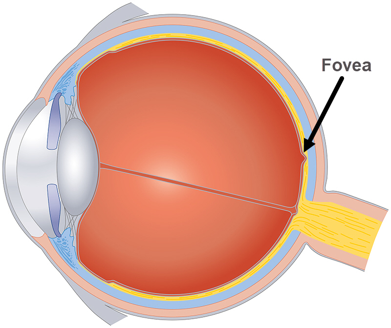
The fovea is a shallow depression at the very center of the macula. The macula is located inside the retina.
Also Known As
- Fovea centralis
- Macula lutea
Anatomy
The fovea measures about 1.5mm horizontally. The depression in the macula occurs when retinal neurons get displaced. The curved wall arising from the depression is called the clivus which gradually slopes to the foveola (the floor).
In the depression are two photoreceptors, rods and cones. Rods provide night vision and aid in visual orientation. They are absent in the fovea.
Cones are densely concentrated in the fovea. There are between 199,000 to 300,000 cones per square millimeter with a total of about 6 million cones in the fovea. The cones are tightly packed with the outer areas elongated so that they appear like rods. The rest of the cones are spread across the remaining part of the retina. The way the layers of the fovea are spread allows light to fall directly on the cones.
Rods and cones convert light to neural signals which are important to the nervous system.
Function
The fovea provides the sharpest, clearest, visual acuity. Cones are responsible for the detailed vision required for activities like reading, driving or seeing nearby objects.
Associated symptoms & disorders
Some of the conditions associated with the fovea are as follows:
- A retinal tear which occurs when the vitreous shrinks and tugs at the retina. If the vitreous pulls away too hard, it may tear the retina in one or several areas. The result is retinal detachment. A retinal detachment can lead to blindness if not treated.
- Choroidal neovascular membranes (CNVM) occurs when blood vessels grow below the retina causing damage. When these blood vessels leak, they may cause vision loss.
- Retinoblastoma is cancer of the eye. It occurs when retinal nerve cells grow in number and size ultimately forming a tumor. The disease mostly affects children.
- A viral eye infection of the retina can result in cytomegalovirus retinitis (CMV retinitis). CMV is prevalent among individuals with a weakened immune system.
- A breakdown of the macula results in Stargardt disease. Photoreceptors die off resulting in blurry vision as well as loss of color vision.
- Retinitis pigmentosa is a group of eye problems that affect how the retina responds to light. The disease leads to a gradual loss of vision.
- Retinopathy of prematurity (ROP) occurs in babies born prematurely. Unwanted blood vessels grow on the baby’s retina.
- Diabetic retinopathy or diabetic macular edema involves the macula and affects patients with diabetes.
- Central serous chorioretinopathy occurs when fluids build up in the retina leading to vision distortion.
- Since the fovea is part of the macula, any disease affecting the macula also affects it. The disorders include histoplasmosis, age-related macular degeneration (AMD), macular edema, macular hole, macular pucker, macular telangiectasia, branch retinal vein occlusion (BRVO), central retinal vein occlusion (CRVO), etc.
Diagnosis of associated disorders
Diagnostic tests for fovea-related diseases include:
- Laboratory tests such as blood sugar, cholesterol and checking for blood clotting.
- An Amsler grid for histoplasmosis to establish the presence of dark, blurry or wavy parts in one’s vision.
- Imaging tests like optical coherence tomography (OCT). OCT scans the retina to establish how thick the retina is.
- An eye examination where the eye is dilated using eye drops. Dilation enables observation of the back of the eye.
- Genetic testing which is relevant in retinitis pigmentosa. Alongside genetic testing is visual field testing and electroretinography.
- Fluorescein angiography which seeks to establish the presence of blood vessel leakage. A yellow dye is injected into an individual’s vein and as it moves through the blood vessels, images are taken.
Treatment of associated disorders
Conditions like central serous chorioretinopathy require no treatment and will resolve on their own. Researchers are yet to discover a cure for Usher syndrome and retinitis pigmentosa. Patients use low-vision devices and techniques to assist them with vision.
Vascular endothelial growth factor (VEGF) is a chemical responsible for the growth of abnormal blood vessels below the retina. Anti-VEGF drugs may treat some of the diseases related to fovea like CNMV. These medications block the chemical to slow the leakage. Steroids are also injected into the eye to help in reducing macula edema (swelling).
Antiretroviral therapy, Ganciclovir and other antiviral medications can treat conditions occasioned by a weakened immune system.
Various forms of laser surgery can be used in the treatment of some of the diseases associated with the fovea or retina. Thermal laser surgery kills abnormal blood vessels and prevents more growth, bleeding or leakage. Photocoagulation and cryopexy may treat retinal tears by sealing the retina to its original position. Scleral buckle and pneumatic retinopexy help push the tear back into place.
Vitrectomy involves the removal of the vitreous gel and replacing it with a gas bubble to treat a retinal detachment.
Scientists are yet to find treatment for some diseases like retinitis pigmentosa. Taking vitamins, including vitamin A palmitate, have been shown to relieve the symptoms. Patients with dry AMD may also take vitamin therapy or nutritional supplements. For Stargardt disease, wearing sunglasses can assist in protection against ultraviolet rays.
Cancer treating therapies are administered for retinoblastoma. They include chemotherapy, radiation, laser, cryotherapy, thermotherapy and enucleation (removal of the eyeball).
