Introduction
The human eye works like a digital camera with complex structures and functions. To enable vision, the eye will adjust itself to the amount of light coming in. It will focus on objects which are close by or far.
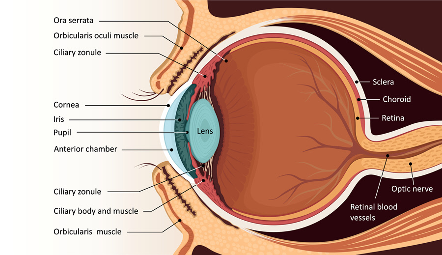
Anatomy
- The orbit comprises of the eyeball, blood vessels, nerves, muscles and a place for tear drainage. Every orbit is shaped like a pear and it contains a number of bones.
- The eye is located inside the orbit with six extraocular muscles attached to it. These muscles are attached to the sclera, the white part of the eye. The extraocular muscles enable the up and down movements of the eye. They also move the eye side to side and rotate the eye.
- The conjunctiva is a clear membrane which covers the surface of the eye. It also covers the inner surface of the eyelids.
- The eye contains the tear film which has three layers: the lacrimal gland, the meibomian gland and tear duct. The lacrimal gland produces the watery part of the tears while the meibomian gland produces oil that forms part of the tear film. Tears lubricate the eye as well as drain through the tear duct.
- The clear, transparent, covering in the front of the eye is the cornea. Located behind the cornea is the anterior chamber which houses the aqueous humor. The eye is always producing the aqueous humor which in turn drains from the eye through the drainage angle. Draining maintains intraocular pressure (IOP).
- The iris is located behind the anterior chamber. The iris is the colored section of the eye with the pupil in the middle. The muscles in the iris aid in controlling the quantity of light that reaches the back of the eye by widening or narrowing.
- The light coming in falls on the cornea which helps to focus it on the retina. After entering the cornea, light travels through the pupil.
- The aqueous humor is produced in the posterior chamber before flowing into the anterior chamber through the pupil. It then drains out through the drainage angle.
- The lens sits directly behind the pupil. The capsule holding the lens is attached to the zonules (small fibers). These zonules suspend the capsule from the wall of the eye.
- When the lens alters its shape, it focuses light onto the retina. The ciliary muscles cause the lens to thicken so that it can focus on nearby objects. The lens can also become thin to focus on far-away objects.
- In between the lens and the back of the eye lies the vitreous cavity which is filled with vitreous humor. Vitreous humor nourishes the inside of the eye and assists the eye to keep its shape. Light entering the eye passes through the vitreous humor onto the retina.
- The retina is sensitive to light and is found at the back of the eye. Inside the retina is the macula and the peripheral retina. The macula provides clear, sharp vision while the peripheral retina provides side vision. The retina sends light through the optic nerve to the brain. The nerve fibers in the optic nerve transmit the light impulses from the retina to the visual cortex in the brain.
- The retina also has special cells, photoreceptors, which change light into energy that is transmitted to the brain. Rods and cones are two types of photoreceptors. Rods perceive black and white and aid in night vision while cones perceive color and give central vision. Inside the retina are blood vessels which nourish it.
- The choroid lies between the retina and sclera and supply it with blood vessels.
Function
A healthy eye functions to provide clear vision. In order to see, light is focused on the cornea with the iris adjusting the amount of light entering the eye. Through accommodation, the lens assists the eye to focus on nearby objects. When the light reaches the retina, it will convert the images into electric signals and transmit them to the visual cortex through the optic nerve.
Associated symptoms & disorders
Any malfunction, disease or disorder of the eye may affect vision. Normal vision involves all the parts of the eye working well. The disorders or diseases may range from minor conditions like eyelid twitching which causes no problems with vision to trachoma that can lead to blindness. Some diseases like bacterial conjunctivitis are caused by a bacteria while a virus causes viral conjunctivitis.
Certain conditions may be congenital such as a coloboma in which a child is born without certain tissue in or around the eye.
Corneal malfunctions or diseases can give rise to conditions like keratoconus, Fuch’s dystrophy, pterygium, etc.
Retinal malfunctions can lead to macular hole, retinal detachment and age-related macular degeneration among others.
Diagnosis of associated disorders
Diagnosis will depend on the eye disease. Generally, an eye care professional may conduct some or all of the following:
- A complete medical history
- Visual acuity test
- Visual field testing
- Color vision testing
- Blood tests and cultures
- Tonometry to check for intraocular pressure
- Ophthalmoscopy to check the back of the eye
- Slit lamp examination which uses high magnification
- Imaging tests such as computed tomography and magnetic resonance imaging
Treatment of associated disorders
Treatment depends on the disease. Some diseases like a stye will resolve on their own. To help with certain symptoms, the doctor may prescribe eye drops and ointments to reduce inflammation, pain and redness in the eye.
Some like bacterial conjunctivitis need antibiotics. Intravitreal steroids and anti-VEGF eye injections can treat retinal diseases such as retinal vein occlusion. Antifungal and antiviral drugs may be used to treat retinitis.
Some cases like cataracts require surgery while others such as posterior capsulotomy are treated by use of laser surgery.
Other like chalazion will need warm compresses.
Refractive diseases are often resolved by use of eyeglasses or contact lenses.
