Introduction
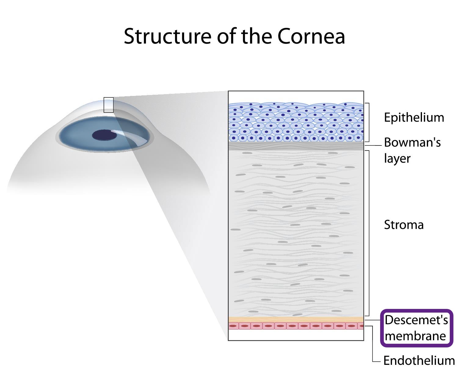
The Descemet membrane is the thin layer of tissue that forms the central connective tissue of the cornea. It consists of collagen and is a specialized basement membrane of endothelial cells. It lies between the corneal stroma and endothelium (the endothelial cell layer).
The DM is the basement tissue for the endothelial cell layer. It is produced by a layer of epithelial cells which form the corneal endothelium. The endothelium sits at the rearward layer of the cornea. Descemet's membrane thickens with age.
Also Known As
- DM
- Descemet's Layer
- The membrane of Demours
- The lamina elastic posterior
- The posterior elastic lamina
- The posterior limiting lamina
Anatomy
The Descemet membrane consists of two distinct layers:
- A posterior non-banded layer (PNBL) which is an amorphous assortment of an extracellular matrix. It lies between the anterior banded layer (ABL) and the corneal endothelium.
- The ABL consists of well arranged collagen fiber bundles. It is situated between the PNBL and the corneal stroma.
Function
The Descemet membrane and endothelium make up the posterior cornea.The cornea plays a critical role in the visual pathway. It acts as a window and the eye's outermost lens that controls incoming light and focuses it onto the retina. It accounts for between 65 and 75 percent of the total focusing power of the eye. The incoming light strikes the cornea which refracts or bends it onto the eye lens.
Descemet’s membrane and endothelium are vital for nourishing the corneal stroma and maintaining the transparency or clarity of the cornea. The clarity and the curvature of the cornea are essential in maximizing visual potential. The DM helps to keep the phenotype and function of corneal endothelial cells which control the amount of fluid in the cornea. It may also support the regeneration of the endothelial cells after a corneal endothelial injury.
Associated symptoms & disorders
Various problems may affect the Descemet membrane including:
- Descemet’s membrane detachment can occur after eye surgery
- Descemet folds linked to corneal edema and corneal inflammation because of endothelial dysfunction from infiltrations, infections or after surgery
- Tears of Descemet's membrane may happen in birth trauma, congenital glaucoma, keratoconus, Terrien's marginal degeneration, keratoglobus, or accidental and surgical trauma.
- Descemet's membrane may also be affected by copper deposition in patients with liver diseases such as Wilson's disease. It can lead to eye discoloration (formation of Kayser-Fleischer rings).
- Fuchs dystrophy, a hereditary condition, may also damage the Descemet’s membrane. In this disorder, the DM becomes extra thick. It causes the endothelium to fail in its function of pumping water out of the membrane and the stroma. The cornea also thickens and clouds due to the interruption of the flow of fluids and nutrients between the cornea and the other parts of the eye.
- Corneal dystrophies are a group of rare hereditary disorders. They cause abnormal material build up in the cornea. It causes swelling of the cornea and can lead to eye discomfort, cloudy vision and permanent vision loss.
Diagnosis of associated disorders
The eye care professional can diagnose Descemet’s membrane disorders based on the patient’s medical history and slit lamp exam of the eye.
Treatment of associated disorders
The treatment method depends on the nature of the disorder.
The DM detachment can be a devastating complication after cataract surgery. Persistent, extensive detachments can affect the sharpness of vision. In severe cases, penetrating keratoplasty surgery (PKP) is the best option for restoration of sight. PKP also referred to as corneal grafting, is a complete corneal transplant. The procedure retains the peripheral cornea and replaces all the damaged or diseased corneal layers with a donor cornea.
Surgery can reverse Descemet’s membrane damage caused by Fuchs dystrophy. Severe damage to the DM may need a corneal transplant. The surgeon removes the damaged membrane and transplants or grafts a new Descemet’s layer obtained from a donor. In most cases, the donor membrane survives and reverses the corneal deterioration.
Descemet membrane endothelial keratoplasty (DMEK) can be used to treat corneal dystrophy. It is a partial-thickness cornea grafting procedure which involves selective removal and replacement of the Descemet membrane and endothelium. DMEK can improve donor corneal graft survival, endothelial cell loss and visual acuity.
The treatment of Descemet folds involves addressing the underlying cause of ocular inflammation. It may include topical antibiotics for infection, topical steroidal, nonsteroidal and osmotic agents.
