Introduction
The cornea is the transparent, outermost layer of the eye. It is tissue with the appearance of a dome. It covers the iris, pupil and anterior chamber. The cornea measures about 12 mm (0.5 inches) in diameter with a center thickness of about 550 microns. It forms a protective window that allows light to enter and focus on the retina. The cornea is responsible for two-thirds of the total optical power of the eye.
For good vision, the cornea must remain clear. It is the reason the cornea has no blood vessels. Instead, it has nerves making it very sensitive to touch, chemicals and temperature. The tears and aqueous humor provide the cornea with oxygen and nourishment.
The curvature of the cornea is spherical but can change with age. The irregular shape of the cornea causes such defects as astigmatism.
Anatomy
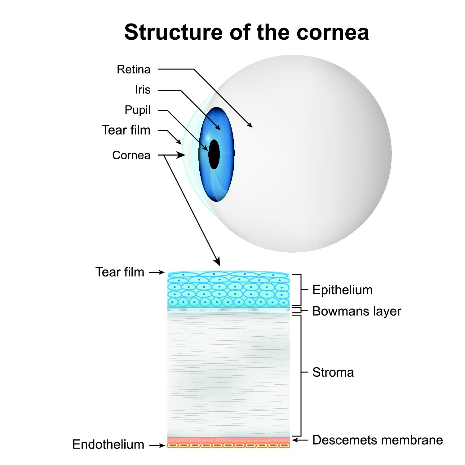
The cornea comprises of five layers:
- The epithelium – The outermost layer of the cornea filled with thousands of nerve endings. The epithelium contains the basement membrane where cells attach and organize themselves. It protects the eye from foreign bodies and infections.
- Bowman’s membrane – It is found in the epithelium. It is the layer at the back of the basement membrane consisting of collagen and protein fibers. An injury to Bowman’s layer may cause a scar which can lead to vision loss.
- Stroma – The stroma lies behind Bowman’s layer. It is the most solid layer of the cornea. The stroma is made up of water and collagen, the reason for the cornea’s shape, elasticity and strength.
- Descemet’s membrane – Tissue located behind the stroma. It fights off infection and injuries.
- Endothelium – It is the thin, innermost corneal layer. Endothelium helps to keep the cornea transparent. Usually, fluid leaks into the stroma from the eyes. The endothelium will push the surplus liquid out of the stroma. This action prevents opacity of the cornea.
Function
The cornea performs the following tasks:
- Protects the eye from foreign bodies and infections
- Helps to focus the light that comes into the eye. The cornea bends light onto the lens. The lens will then refocus that light onto the retina.
- Screens out harmful ultraviolet (UV) rays
Associated symptoms & disorders
The following may affect the cornea:
- Injuries to the cornea – Minor injuries such as scratches (corneal abrasion) heal on their own. Significant injuries can lead to corneal scarring and vision impairment
- Allergies – The most common allergen is pollen. The symptoms will often go away without medical intervention
- Keratitis – An inflammation of the cornea (corneal ulcer). A minor injury or overuse of contact lenses can cause non-infectious keratitis. Virus, bacteria and fungi produce infectious keratitis. The inflammation often goes away with antibacterial, antifungal and steroid treatment
- Dry eye – A condition where the eye has less or poor quality of tears. The eye is not able to lubricate its surface
- Corneal dystrophies also affect the cornea. In corneal dystrophy, some parts of the cornea may lose their normal vision. These dystrophies are hereditary and occur in both eyes. They also progress slowly. Healthy people are also at a risk They include:
- Keratoconus – A progressive thinning and bulging of the cornea. The bulge then forms an abnormal curvature (cone). Keratoconus can cause blurred vision, hyperopia, myopia, double vision, increased sensitivity to light and astigmatism
- Fuchs’ dystrophy – A slow-progressing disease. The cells in the endothelium gradually deteriorate. The result is the thickening of the cornea where vision becomes blurred
- Lattice dystrophy - The accumulation of an abnormal protein fiber called amyloid. It impairs vision with time
- Map-Dot-Fingerprint dystrophy (epithelial basement membrane dystrophy) - Occurs when the tissue on the basement membrane develops folds to impair vision.
- Other diseases include:
- Ocular herpes – A recurring viral infection caused by the herpes simplex virus. It can also be sexually transmitted. The inflammation can spread into the cornea causing stromal keratitis, a more severe infection.
- Iridocorneal endothelial syndrome (ICE) – Glaucoma, swelling of the cornea and changes in the iris will occur.
- Pterygium – A triangular-shaped growth on the cornea. Pterygium is harmless unless it grows too large.
- Stevens Johnson syndrome (erythema multiforme major) - A skin disorder affecting the eyes. It may give rise to corneal blisters, erosions and holes. It can lead to loss of vision.
Diagnosis of associated disorders
The eye care professional will check for a problem with the cornea using the following:
- A detailed patient history
- A slit lamp and an advanced diagnostic technology called corneal topography
- Dilation of the eyes to examine the retina to check for early signs of disease
- Testing for corneal pressure
- Measuring the thickness of the cornea
- A corneal cell count to determine the shape, size and number of cells
Treatment of associated disorders
Antibiotics and steroid medications may treat cases involving injuries, allergies and so on. Rigid gas-permeable (RGP) contact lenses are used to reshape the cornea and improve vision. Dystrophies and other diseases may require surgery procedures like:
- Laser surgery - Phototherapeutic keratectomy (PTK) makes use of UV light. Laser reshapes and restores the cornea
- Corneal transplant surgery – Some corneal transplants involve replacing only some parts of the cornea with tissue from a donor (lamellar keratoplasty). In penetrating keratoplasty (full thickness transplant), the entire cornea is transplanted
- Anterior lamellar keratoplasty - This procedure replaces the damaged tissue in the stroma with healthy stroma from a donor
- Endothelial lamellar keratoplasty – It replaces the diseased endothelial tissue with healthy endothelium from a donor
People who fail with tissue implants can have an artificial cornea in a procedure called a keratoprosthesis (KPro). The Boston type-1 KPro is an example.
