Introduction
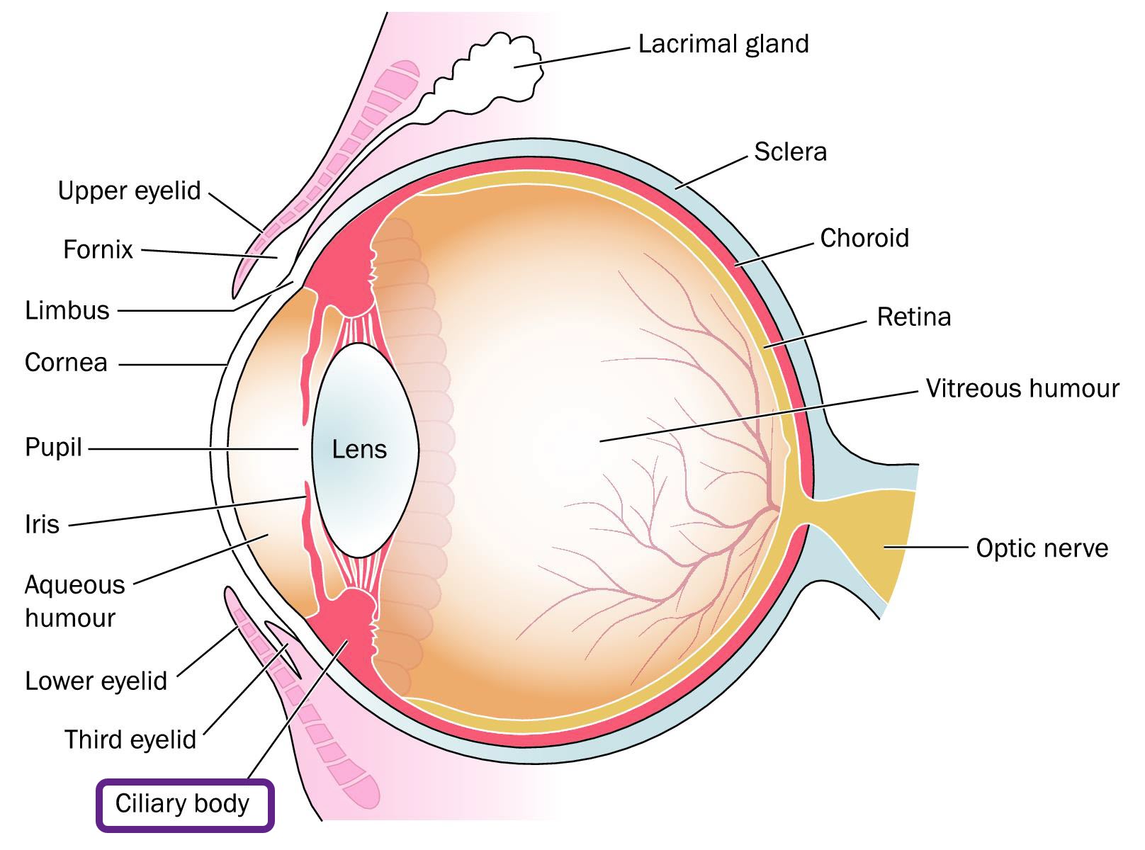
The ciliary body is a muscular tissue located right behind the iris. It divides the vitreous body from the posterior chamber. Together with the iris and choroid, the ciliary body makes up the uvea.
Anatomy
The ciliary body is ring-shaped. It is made up of different parts which play different roles. They include:
- Ciliary muscle – The muscle is situated close to the sclera. It has different fibers arranged either longitudinally, obliquely or circularly. It is responsible for changing the shape of the lens to focus on objects. The muscle is attached to the capsule of the lens by the zonular fibers/ligaments.
- Epithelium – The ciliary body is made up of a double layer, known as the epithelium. The outer layer is an extension of the retinal pigmented epithelium (RPE). The inner layer extends from the neural tissue in the area of the retina. Unlike the outer epithelium, the inner epithelium is not pigmented. It is a transparent layer that comes into contact with the vitreous body.
- Connective tissue – The inner connective tissue is located between the ciliary epithelium and the muscle. It is thinner and more vascularized in the anterior portion of the ciliary body than the posterior portion.
- Ciliary processes – They are formed by the inward folding of numerous layers of the choroid. It is found in the inner non-pigmented epithelium.
- The ciliary body is divided into two portions:
- Pars plicata – This is the anterior part that makes up a third of the whole ciliary body. About 60 ciliary processes are contained in this portion. The pars plicata is responsible for secreting the aqueous humor into the posterior chamber of the eye. It can also be referred to as the ciliary crown. The inner layer of this part is thick and highly vascularized.
- Pars plana– The posterior part of the ciliary body makes up the remaining two thirds. It does not produce any fluid. During surgery or diagnosis, devices are inserted to the eye through the pars plans. The inner epithelium is thin and less vascularized compared to that of pars plicata.
Function
The ciliary body has two main functions which are:
- Accommodation – When the ciliary muscle relaxes, the suspensory ligaments become taut and the lens flattens. This allows for the eye to focus on objects that are far. When the muscle contracts, the ligaments relax and the lens becomes spherical. The release enables the eye to adapt to near vision.
- Production of aqueous humour – The ciliary processes contained in the epithelium produce the aqueous humor. The fluid fills up the chambers and also nourishes the tissues of the eyes.
Associated symptoms & disorders
The diseases that are associated with the ciliary body are such as:
- Glaucoma – The disease is characterized by high intraocular pressure (IOP). The ciliary body is responsible for the production of the aqueous humor. The fluid raises the pressure when secreted in high amounts or when not properly drained. Glaucoma can lead to blindness if not detected and treated early.
- Ocular hypertension – This is a condition where the pressure in the eye is higher than normal but has not yet caused progression to glaucoma. IOP levels above 21 mmHg are considered high.
- Intermediate uveitis – This is inflammation that primarily involves the anterior vitreous and the ciliary body. It is characterised by pain and swelling.
- Iridodialysis – This is the tearing of the iris from its attachment to the ciliary body. It can occur due to trauma/injury. Usually, hyphema follows after the detachment. This is where blood from the ciliary body moves to the anterior chamber.
Diagnosis of associated disorders
Tests that can be made to analyse for disorders of the ciliary body include:
- Slit lamp exam – The test involves use of a slit lamp and a special lens to view the inside structure of the eye. Swelling can indicate uveitis or iridodialysis. Clots of blood can indicate hyphema.
- Fundoscopic exam - An ophthalmoscope is used to inspect the back of the eye. The test can also show swelling and other signs in conditions such as uveitis.
- Tonometry – The test measures the eye pressure. Levels that are too high could indicate ocular hypertension.
- Visual field test – After a tonometry that shows high IOP, a visual field test can help determine whether one has glaucoma. Loss of side (peripheral) vision is common in patients with glaucoma.
- CT scan – In some instances, a CT image can be used to assess problems affecting the ciliary body.
Treatment of associated disorders
Most treatments are aimed at reducing the production of aqueous humor or improving the drainage. Others treat the pain or swelling associated with the disease/disorder. The treatments may include:
- Beta blockers – These are prescribed in glaucoma to decrease aqueous fluid secretion. They include Betaxolol and Timolol
- Carbonic anhydrase inhibitors – They are also given to glaucoma patients to inhibit production of the aqueous humor.
- Alpha-adrenergic agonists – These drugs improve drainage as well as decrease fluid production. They include Apraclonidine and Brimonidine.
- Surgical repair – In some instances, surgical repair may be needed to open/widen the drainage angle.
- Corticosteroids – They are prescribed to reduce inflammation conditions such as uveitis.
- Pain relievers – Where there’s pain such as in iridodialysis, drugs that alleviate pain can also be issued.
For some conditions, the eye doctor may decide to monitor the progression without issuing any medication. In iridodialysis, for instance, a little bed rest may help in managing the condition.
