Introduction
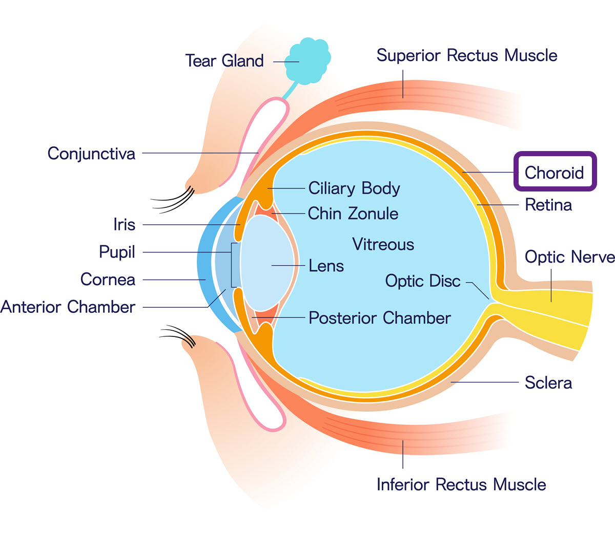
The choroid, the iris and the ciliary body are the three components of the uveal tract. The choroid is a pigmented vascular layer shaped like a rounded wine glass. It lies between the white outer layer (the sclera) and the inner layer of nerve tissues at the back of the eye (the retina). The choroid is a thin membrane filled with blood vessels that supply nutrients and oxygen to the eye.
Healthy eyes and good vision depend on enough blood supply, which means the choroid must be as healthy as possible at all times. When the choroid or the surrounding area is infected, the macula and optic nerve may suffer. When the macula and optic nerve are affected or compromised, one may experience a severe decrease in vision and even total blindness.
Also Known As
- Choroidea
- Choroid coat
Anatomy
The choroid has four different layers:
- Bruch's membrane – It is the central area
- Sattler's layer - It has the medium size blood vessels
- Haller's layer – It is the outermost layer and has the larger blood vessels
- Choriocapillaris – This area has the tiny blood vessels (capillaries) that distribute blood
Function
The primary task of the choroid is to supply oxygenated blood to the outer layers of the retina. Other functions include:
- Assisting in the control of intraocular pressure
- The choroidal blood flow may regulate retinal heat
- It may serve to protect the choroidal blood vessels against light toxicity
- The choroid may be involved in the growth of the sclera through cells that secrete particular substances
- The dark-colored melanin pigment limits reflections within the eye and absorbs the light that could degrade vision
- The choroid can change its thickness and bring the photoreceptors into the plane of focus; these changes can cause the retina to move forward and back
Associated symptoms & disorders
The choroid may be affected by various conditions such as:
- Choroidal nevi – It is a build up of pigmented or nonpigmented cells in the choroid
- Chorioretinitis - It is the most common disease of the choroid. Often, this inflammation produces a blurry vision and floating dark spots. It usually affects young children and people with Herpes Simplex Virus.
- Choroidal dystrophies – It is a group of inherited diseases that include pigmented paravenous retinochoroidal atrophy, choroideremia, central areolar choroidal dystrophy, diffuse choroidal atrophy and gyrate atrophy. These diseases can cause severe loss of vision.
- Choroidal detachment and hemorrhage - The choroid can detach from the sclera causing the accumulation of blood or fluid. The rupture of choroidal vessels can cause a leakage in the choroid or space above the choroid. This condition is scarce and is associated with blunt trauma, eye surgery, inflammatory disease, uveal effusion syndrome and intraocular tumors.
- Choroidal rupture - It occurs when there is a complete tear in the retinal pigment epithelium, Bruch's membrane and the choroid. It happens as a result of blunt eye trauma like being hit with a fist. Often, choroidal ruptures involve the center of the retina (the macula) which controls full vision abilities. A rupture can cause acute preretinal, retinal or subretinal hemorrhaging.
Diagnosis of associated disorders
The eye care professional can diagnose choroidal diseases and disorders through a comprehensive eye exam. The tests may include:
- CT
- MRI
- B-Scan ultrasound
- Ultrasonography
- Fundus examination
- Fluorescein angiogram
- Optical coherence tomography
Treatment of associated disorders
The mode of treatment depends on the nature and extent of the condition.
- Chorioretinitis – The doctor can prescribe antibiotics and corticosteroids
- Choroidal rupture – Surgery is needed to repair the damage and medication may be prescribed to help in the healing process
- Choroidal dystrophies – These are genetic disorders with no definitive or standard treatment. The eye doctor may employ symptomatic treatment to address the signs and symptom.
- Choroidal detachment and hemorrhage - Treatment consists of eye pressure lowering eye drops, topical steroid eye drops and cycloplegic eye drops. Surgery may also depend on the nature of the detachment. Operative choroidal hemorrhage occurs during surgery and is handled in the operating room.
- Choroidal nevi – Most choroidal nevi don’t need any treatment and only require monitoring. The eye doctor photographs the area of concern and tracks it. If the choroidal nevus appears elevated, has an unusual shape or has orange pigmentation, it may become a malignant choroidal melanoma. Aggressive treatment is required.
