Introduction
Retinoscopy is a technique that examines the retina to provide an objective assessment of the refractive error. It enables the eye professional to ascertain if there is a need for vision correction. The test is part of a comprehensive eye exam conducted to check the state of the eyes and focus on eye care.
If a patient has a problem with their eyesight or is experiencing a change in vision, they might be suffering from a refractive error or an eye disorder. The eye doctor may conduct a retinoscopy to examine eye health. It is an easy, quick, and reliable test that needs minimal effort or response from the patient.
Also Known As
Skiascopy
Purpose
A retinoscopy is an initial procedure to diagnose the refractive error of the eye. The assessment is made during a routine eye exam and is conducted at the eye clinic or the eye doctor's office by:
- a technician
- an optometrist
- an ophthalmologist
It helps to determine whether a patient is suffering from emmetropia, nearsightedness (myopia), farsightedness (hyperopia), or astigmatism. It also helps in detecting eye diseases and defects such as macular degeneration, retinal vessel occlusion, retinal detachment and injuries in the eyes.
A retinoscopy exam is an objective procedure that requires little cooperation from the patient. It is therefore useful in determining the refractive error in infants, young children and the elderly.
The procedure also helps to diagnose vision problems in developmentally delayed adults. It is useful for people with a disability or whose behavior limits their ability to respond or cooperate with other refraction techniques.
At times, a retinoscopy may be the only eye exam needed; mainly where a patient has emmetropia and will have a better vision without eyeglasses or contact lenses.
However, where refractive errors are detected, the test is often followed by other subjective procedures. These subsequent tests help determine the accurate corrective lens prescription the patient needs for clear vision and any other required medications.
Preparation & Expectation
It is a quick and pain-free test although one may experience some tearing or watery eyes when exposed to the light beam from the measuring instrument, the retinoscope.
There aren't any eating restrictions before this test. However, for an accurate result of the exam, the eyes must be free from external products or devices such as contact lenses. The eyes may be diluted before the exam.
Generally, children's eyes are dilated for the procedure because it is easier to observe how light bounces on the back of the eye when the pupil is enlarged. Also, the drops temporarily restrict the eye's accommodative ability or focus, which allows the doctor to make a more accurate assessment of the refractive error. In some cases, children may need general anesthesia.
Procedure
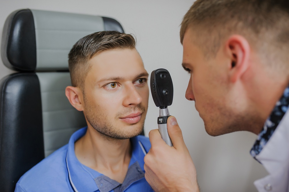
The handheld retinoscope can automatically detect a vision problem. It consists of a mirror and a condensing lens and projects a ray of light into the eye. The eye professional will move the light beam vertically and horizontally to examine the image from different angles.
So during the exam, one has to sit upright to reduce disturbances that can be caused by unnecessary body movements. The examiner will ask the patient to look straight at the measuring instrument to allow the light to pass through the eye pupil. Better images are observed when the eyes are diluted.
The doctor will assess the light reflecting off the back of the eye and diagnose eye health. He/she will then introduce different lenses in front of the eye. As the doctor inserts lenses with different power there is a corresponding shift in the pattern and direction of the reflection.
The doctor will keep changing the lenses until he/she gets the lens power that indicates a refractive error. The procedure is done for both the eyes, one at a time, and takes about 10 minutes to complete.
A child will have to look at the retinoscope for a short period that varies from several seconds to a few minutes. Often, children, including infants, can look directly at the instrument long enough for a successful retinoscopy without anesthesia.
Modern high-tech equipment such as autorefractors, make the retinoscopy exam much more comfortable and faster. They take measurements automatically in a matter of seconds.
Outcome
The most important aspect of this exam is the assessment of how well the eyes focus on the light beam. It is a measure of focal length i.e., it determines the exact angle at which the ray of light refracts off the back of the eyes.
The measure is what lets the doctor know how well the eyes can focus and enables them to determine whether one needs eyeglasses. The results of the test also provide information about risk factors for other eye disorders.
Individuals suffering from nearsightedness are at an increased risk of having severe other eye diseases, such as glaucoma and retinal detachment. Patients with severe farsightedness have significant chances of developing eye disorders like accommodative esotropia and amblyopia.
A small group of children having high farsightedness and crowded anterior segment have a higher risk of developing angle-closure glaucoma.
Risks & Complications
Retinoscopy is a safe procedure with no known risks.
