Introduction
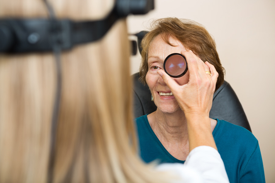
Ophthalmoscopy is an eye examination which allows an eye specialist to look at the back of an individual’s eye. Often, this test is part of a routine eye exam by an optometrist or ophthalmologist.
Ophthalmoscopy is conducted through a hand held device known as an ophthalmoscope. The instrument is of two types:
- A panoptic which looks like a telescope.
- The standard head which is the traditional ophthalmoscope.
The eye doctor may also use an instrument called a slit-lamp.
Also Known As
- Funduscopy
- Retinal examination
Purpose
The eye care professional can use ophthalmoscopy to check for eye diseases and other disorders. These eye conditions include:
- Retinal tear or detachment
- DiabetIc retinopathy
- Damage to the optic nerve
- Glaucoma
- A viral retina infection known as cytomegalovirus (CMV) retinitis
- Macular degeneration
- Melanoma
Types
- Direct ophthalmoscopy
The professional uses the ophthalmoscope to shine light into the patient’s eyes. The ophthalmoscope also has lenses that allow the specialist to examine the patient’s eyes.
- Indirect ophthalmoscopy
This eye test uses the standard head ophthalmoscope. This method provides the eye professional a better look at the retina compared to the other methods.
The method can also be combined with an eye exam technique known as scleral depression. The combination enables the ophthalmologist to look at the far edges of the retina and detect detachment or tears.
- Slit-lamp ophthalmoscopy
This method uses a slit lamp to shine a high intensity light into the patient’s eyes. It gives the same view as an indirect examination. Yet, it offers greater magnification, bigger images.
Preparation & Expectation
Before conducting the eye exam, the eye professional may use eye drops to dilate the patient’s pupils. Dilation makes the pupils larger and easier to look through.
Patients should be aware that the eye drops may cause sensitivity to light and blurred vision for a few hours. The eye drops may also sting the patient’s eyes a bit and bring an unusual taste in the mouth. Sunglasses can help to protect the patient’s eyes from bright light while the pupils are dilated.
Where a patient is allergic to any medication, it is vital to inform the eye specialist in advance. It is also important to disclose any medication the patient is taking. This includes dietary supplements, prescription medications and OTC medications.
The eye professional should also know if the individual has glaucoma or a family history of the condition.
The patient can’t drive or perform functions that need clear vision for a few hours after the eye test.
Procedure
Before conducting the eye exam, the eye professional may use eye drops to dilate the patient’s pupils. Dilation makes the pupils larger and easier to look through.
Patients should be aware that the eye drops may cause sensitivity to light and blurred vision for a few hours. The eye drops may also sting the patient’s eyes a bit and bring an unusual taste in the mouth. Sunglasses can help to protect the patient’s eyes from bright light while the pupils are dilated.
Where a patient is allergic to any medication, it is vital to inform the eye specialist in advance. It is also important to disclose any medication the patient is taking. This includes dietary supplements, prescription medications and OTC medications.
The eye professional should also know if the individual has glaucoma or a family history of the condition.
The patient can’t drive or perform functions that need clear vision for a few hours after the eye test.The eye tests involve the following:
- Direct ophthalmoscopy
This exam is conducted in a dark room. With the patient seated on a chair staring straight ahead, the eye doctor shines light into the eyes and examines them. The patient may be asked to move the eyes in certain directions.
- Indirect ophthalmoscopy
The patient sits in a reclining position or lies down on a coach. The specialist shines light into the patient’s eye while holding it open. The patient may be asked to look in certain directions during the exam. The eye professional may also use a small blunt probe to apply some pressure to the patient’s eye.
- Slit-lamp ophthalmoscopy
The patient sits with the chin and forehead resting on the device called a slit lamp. The eye care professional shines a bright light into the patient’s eyes and examines them with a microscope. He/she may ask the patient to move the eyes and use fingers to open the patient’s eye for a better look. They may also use a small blunt probe to apply some pressure to the patient’s eye.
These eye checkups generally take about five to ten minutes.
Outcome
If the optic disc, retina, and blood vessels show no sign of disease or pathology, no further action may be necessary.
Abnormal results, such as spots on the retina or swelling, could be signs of disease.
Risks & Complications
At times, ophthalmoscopy may be a little uncomfortable, but it isn’t painful.
In rare cases, patients may react to the eye drops which may cause:
- Dizziness
- Dry mouth
- Nausea
- Vomiting
- Flushing
- Narrow-angle glaucoma
