Introduction
A retinal tomography is an automated diagnostic procedure which maps the retina’s surface, optic nerve head as well as the posterior segment/back of the eye. The optic nerve is responsible for transmitting unique sensory information for vision processing from the retina to the brain.
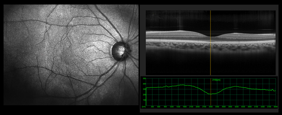
HRT uses a specialized scanning laser ophthalmoscope to take topographic 3-D photographs of the optic nerve. This laser is pointed on the surface of the optic nerve to capture the image. HRT uses the reflection of light from the retina to take pictures of the structures. It takes 32 confocal photos in 1.6 seconds. It then creates an image of the surface topography by producing a 3-D picture.
HRT is more superior when compared to a hand-drawn image or a regular photograph of the eye. This is because it uses software to analyze images taken to a pre-existing directory that contains images of healthy eyes. The software also checks whether the recorded measurements are within the normal range or not. It is also considered more superior because it provides disease progression analysis, which helps give early glaucoma warnings.
Also Known As
- Heidelberg Retinal Tomography (HRT)
Purpose
HRT is an eye exam that must be performed by an eye care specialist to enable precise diagnosis, treatment, and management. HRT is usually conducted after a clinician consultation by:
- An ophthalmologist
- An optometrist
- A technician
The test is widely used to detect glaucomatous visual field changes even before visual field defects can be seen. The test can confirm a suspected case of glaucoma or predict the risk of early glaucoma in patients above the age of 40. Glaucoma, which is caused by high eye pressure, is a leading cause of blindness in people over 60 years of age. Risk factors for developing glaucoma include age, race, family history, eye pressure, and myopia. Blindness caused by glaucoma is irreversible. It is therefore vital to have regular checks conducted.
Glaucoma damages the optic nerve, which is a part of the eye. When a considerate amount of optic nerve cells die, they result in a small ‘cup’ in the eye. This optic nerve damage is known as ‘cupping’. One of the things that eye doctors look out for when conducting HRT is the occurrence and measure of the ‘cup’.
It also provides a more objective method to detect or follow up on the progression of various retina diseases like:
- Age-related macular degeneration (AMD)
This disease affects the macular, which is part of the retina. AMD tends to appear as one progresses in age, especially in patients over 60. People with AMD experience fuzzy, shadowy, or distorted vision.
- Retinal detachment
This is a disorder where the retina separates the blood vessels that provide it with nutrients. Symptoms include flashes, increased floaters, and losing sight in the outer visual field. Total vision loss can occur without appropriate treatment.
- Diabetic retinopathy
This is damage to the retina’s blood vessels caused by a diabetic eye disease (diabetes mellitus). It occurs when there are high blood sugar levels. The eye disease may not present any symptoms and may result in vision loss.
- Macular holes or edema
Macular holes are small breaks, while edema is the fluid build-up, both present in the macular. They can result in blurry central vision.
Preparation & Expectations
Patients who wear glasses or contact lenses are required to know their specific glasses’ prescription. This is because the doctor will dial in these details into the Heidelberg Retinal Tomographer. The procedure does not require the dilation of the pupils, although this depends on the doctor performing the procedure.
HRT is a simple, painless, and non-invasive procedure. The procedure takes only 10-15 seconds for each eye, and because of this, people with special needs can easily get tested.
Procedure
The patient will be asked to sit directly in front of the Heidelberg Retinal Tomographer. The doctor will turn down the lights in the room and ask the patient to take off their glasses/contact lenses. They will then place their chin on the rest and lean their forehead on the headband. The patient will look directly into the instrument with their eyes fixated on the camera right ahead of them. The doctor will direct the patient to blink as usual to keep their eyes from drying out.
The examiner aligns the instrument to be parallel to the patient’s eye, and once in position, the laser will begin scanning the retina. The device will create a surface map of the patient’s retina. It only takes a few seconds. When it’s done, the same procedure is repeated on the other eye.
Outcome
- Normal results
Results are considered positive when the appearance of the optic nerve is healthy. No fluid, blood, or ‘cup’ is observed in the 3-D image.
When the produced HRT 3-D scan shows any abnormalities, it may indicate the presence of:
- Retinal detachment
- AMD
- Macular holes or edema
- Diabetic retinopathy
For patients who have been diagnosed with any of the above diseases/abnormalities, frequent repeat evaluations may be recommended to monitor the progress.
Complications
There are no known complications because the laser used in the procedure is entirely harmless to the patient.
