Introduction
A pupil test is an eye assessment to evaluate the appearance and function of a patient's pupils. It is an essential component of a comprehensive eye exam. The eyes can provide critical information about the state of a person's health. It's the reason why doctors use a variety of techniques to diagnose a patient's eyes.
The black round area in the middle of the colored portion of the eye (the iris) is the pupil. It is an aperture or window which allows light to reach the back of the eye, the light-sensitive spot in the retina. It works like a camera aperture by regulating the amount of light that enters a person's eyes.
The pupil can dilate i.e., expand and become more extensive or constrict, become smaller. The iris muscles respond to external stimuli and regulate the amount of light that flows to the back of the eye. In dim light or a dark environment, the pupil dilates to let in more light and improve the vision. In bright light, it constricts to decrease the amount of light reaching the back of the eye.
One should see a doctor if they notice that the pupils have an unusual appearance. Most noteworthy, one should get immediate medical assistance if they also start to experience dizziness, confusion or severe head pain.
Also Known As
- Pupillary examination
- Pupillary response test
- PERRLA eye examination
Purpose
The pupil test can be conducted at the doctor’s office or eye clinic by:
- an optometrist
- general physician
- an ophthalmologist
A pupillary assessment can help the doctor to diagnose a variety of eye and health disorders ranging from common conditions, such as migraine to retinal and neurological disorders.
A person does not have voluntary control of their pupils; therefore, a pupil test may reveal problems with a patient's autonomic nervous system and other areas of the body. Thus, the pupillary response test is an essential component of the eye and neurological examinations because changes in the patient’s pupils’ reactivity, size, and equality may offer critical diagnostic information.
Preparation & Expectation
Pupil eye exams are quick, noninvasive tests that don't require any preparation.
Procedure
There is a long nerve cord in the body that regulates the pupil. It originates in the brain and moves down to the spinal cord. It travels to the lungs, subclavian arteries, neck, through branches of the mind and along the optic nerve, and ends up at the pupil. An issue along this cord is likely to impact the nerve and lead to changes in pupillary response.
The acronym PERRLA assists doctors in remembering what to check for during the pupil examination. It stands for:
- Pupils- The pupils control light entering the eye by widening and shrinking.
- Equal- The pupils should be of the same size.
- Round- They should be well rounded.
- Reactive- They should react to the external stimuli by responding to the amount of light entering the eyes. It reminds the doctor to assess the patient's pupils' responses to the last two items.
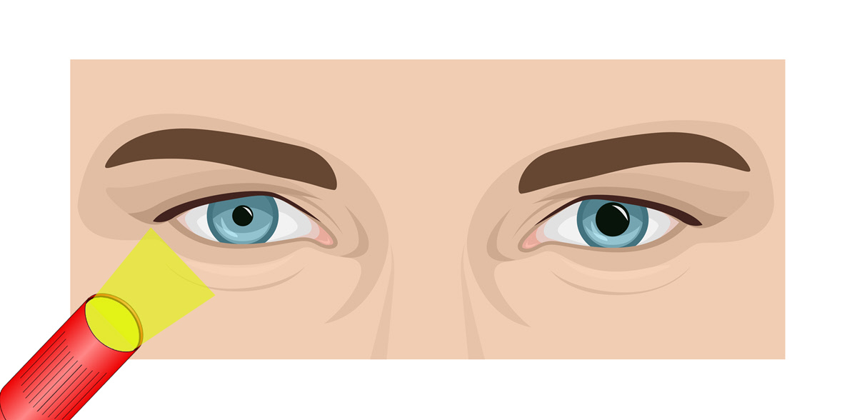
- Light- The doctor shines a light into that patient's eyes and assesses the pupils' reaction.
- Accommodation- The eyes can see objects that are both close up and at a distance. The patient will sit in a dimly lit room, and the doctor will use three procedures to assess the pupil reflexes.
Light response pupil test- It examines the intuitive movement that regulates the patient’s pupil size as it reacts or responds to light. The doctor dim's the lights and asks the patient to focus on a target in the distance. He/she then shines a light into the patient's eyes from each side and carefully watches the pupils to determine whether they are constricting in response to the light and also check for anything unusual about their shape or size.
Swinging flashlight pupil test- The test compares the patient's pupils' reaction to light. In the dimly lit room, the patient is again requested to look at a target in the distance. The doctor will then rhythmically oscillate the light from eye to eye and examine each pupil's response.
Near response pupil test- The test evaluates how the pupil responds to a near object. It is conducted with standard lighting in the room. The doctor will ask the patient to focus on a target far away and then pass a card or small object in front of the eyes. As the patient fixates their eyes on the target, he/she will carefully examine the pupils to ensure that they constrict rapidly as the patient's fixation changes from far to near.
Outcome
The results of a pupil test can uncover many disorders, depending on which portion of the exam was abnormal.
I. Uneven shape or size
The doctor begins the test by checking the pupils for anisocoria. It is an abnormality in which the patient's pupil sizes are unequal i.e., they aren't perfectly round, or have a difference of over 1 millimeter in size. It does not indicate anything abnormal because about 20% of the population has normal anisocoria. However, in some patients, unequal pupil sizes may be a symptom of disease affecting their blood vessels, brain, or nerves.
Conditions that cause unequal pupils include:
- Stroke
- Seizure
- Migraine
- Aneurysm
- Glaucoma
- Brain tumor
- Brian inflammation
- Intracranial hemorrhage
- Brain trauma, such as a concussion
II. Not reactive to light or accommodation
The patient's pupils should remain in the same size or constrict when light is shone on them. If the pupils dilate it may indicate an optic nerve problem, including:
- Glaucoma
- Optic neuritis
- Retinal infection
- Optic nerve tumor
- Optic nerve damage
- Ischemic optic neuropathy
- A hyperactive ciliary muscle
It is important to note that the pupil test results on their own aren't enough to diagnose any disease. They only provide the doctor with a better understanding of what other exams he/she can use to assist in narrowing down the probable causes of the patient's symptoms.
Risks & Complications
The pupil test is a simple eye exam without risks.
