Introduction
Optical coherence tomography is an imaging technique used in clinical diagnostics to check internal structures. It is a light-based imaging technology which uses optical interferometry. Light reflected from a structure under examination combines with reference light to generate interference signals that create 2D and 3D images.
Optical coherence tomography can use longer light wavelengths for deeper penetration. It makes the technology suitable for non-destructive sample testing and non-invasive biomedical imaging. OCT allows non-contact, non-invasive and real time measurements for 3D visualization.
It is best suited for clinical biomedical imaging and biophotonics applications in optometry and ophthalmology. It allows optometrists and ophthalmologists to get detailed information about the internal microstructures. These include the retina, macula, optic nerve and choroid.
The technique gives ophthalmologists and optometrists high resolution cross sectional images of the inner eye. It is among the latest technological advances which allow eye care professionals to analyze the anatomic structure of the retina and optic nerve disc. The distinctive layers of the retina are viewed, mapped and their measurements taken for a specific diagnosis.
Also Known As
Optical Coherence Tomography
Purpose
Optical coherence tomography is used to:
- Check the retina and the retinal structures
- Assess retinal abnormalities or defects
- Make detailed measurements of the optic nerve head and the retina
It is now the standard clinical imaging procedure for screening and diagnosis of many eye conditions including:
- Glaucoma
- Retinal Tear
- Macular holes
- Macular pucker
- Vitreous traction
- Choroid melanoma
- Retinal detachment
- Diabetic retinopathy
- Optic nerve disorders
- Macular degeneration
- Pre-retinal membranes
- Cystoids macular edema
- Central serous retinopathy
- Preretinal macular fibrosis
OCT also helps doctors to develop a treatment plan and track the outcomes of the procedures over time.
Preparation & Expectation
Optical coherence tomography is a quick and painless medical exam. It is a non-contact and non-invasive scan of the inner eye microstructures.
Dilating eye drops will be applied into the patient’s eyes if:
- The pupils are very small
- The professional needs an image of a very specific area
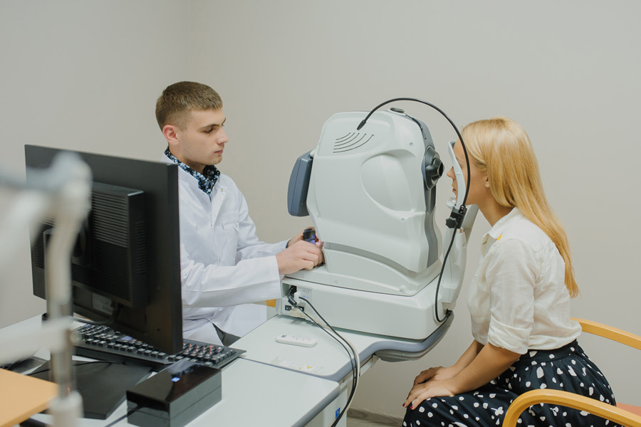
Procedure
The exam is performed in the eye doctor’s office. It takes about five to ten minutes. The patient sits in front of the OCT machine and the technician calibrates it. The instrument projects a beam of light onto the retina. Its specialized electronic system detects, gathers, processes and stores the echo signals. It uses the generated patterns to create cross sectional pictures of the retina and conducts the necessary measurements. The instrument displays the results on a screen for evaluation and analysis.
Outcome
Optical coherence tomography is sophisticated technology. It produces very high resolution 3D pictures of the inner eye structures such as the cornea, iris, retina, eye lens, vitreous and optic nerve head.
The snapshot in time allows the eye doctor to isolate and differentiate the layers of the retina. It enables the doctor to evaluate the vitreoretinal interface and to detect retinal swellings. He/she can identify retinal and optic nerve diseases.
If the retina, the retinal structures and optic nerve disc are of normal size and shape, it means the eyes are healthy.
If the scan reveals defects such as swellings or pits, it is an abnormal result. It may indicate the presence of a disease.
Optical coherence tomography shows the exact layer of the retina where the fluid is building up and causing retinal swelling. It enables the doctor to decide where treatment is necessary. He/she can also assess whether retinal treatments such as injections or laser therapy can decrease the accumulation of fluid or the swelling in the retina.
The instrument uses the stored results to track the healing or progression of the disease. The doctor can compare past and present results to find small changes over time.
Risks & Complications
Optical coherence tomography is approved for use in the US. It is an advanced technology with minimal risks or complications for patients. OCT presents no radiation risk because it doesn’t use X-ray technology. The light that is projected into the patient’s eye is safe. Where there may be a risk to the patient, the instrument is designed to end the test before any damage occurs.
