Introduction
Indirect ophthalmoscopy is an essential method of fundus examination. The fundus is an area at the back of the eyeball and comprises blood vessels, retina, and the optic nerve. The retina is the light-sensitive spot that recognizes light and images, and the optic nerve is the cable that carries the signals to the brain. The doctor uses an instrument called an ophthalmoscope, which is a magnifying tool, and a light source to view the structures inside the eye.
Indirect ophthalmoscopy is often included in standard eye tests and routine physical exams to screen for eye disorders. It is particularly essential in cases of a suspected detachment of the retina. The doctor can order the test if a patient has a condition or an illness that affects their blood vessels, like diabetes and hypertension.
Other eye exams might be done along with indirect ophthalmoscopy. These may include:
- Tonometry
- Vision Tests
Also Known As
- Indirect Funduscopy
- Retinal examination
Purpose
Indirect ophthalmoscopy requires excellent skill and specialized tools. It is usually done by:
- Optometrists
- Ophthalmologists
Indirect ophthalmoscopy provides the doctor with a greater depth of focus, a more brilliant picture, and a full field of view. It gives a three-dimensional (3-D) image of the back of the eye, which allows the doctor to have a detailed picture of eye abnormalities. He/she can use to test to screen for eye disorders such as:
- Diabetes
- Glaucoma
- Brain tumors
- Head injuries
- Choroidal lesions
- Damaged optic nerve
- Macular degeneration
- Retinal tear or detachment
- Hypertension or high blood pressure
Preparation & Expectation
The doctor will apply eye drops in the patient's eyes before the test to dilate the pupils. It makes them expand and more accessible for the doctor to look through and get a better view. The drops will take effect in about 20 minutes and keep the patient's eye dilated for several hours. Therefore, the patient will need to wear sunglasses after the test until the drug effects wear off. The patient may also have a blurry vision temporarily after the test and will need someone to take them home. People whose work requires a clear vision should take the rest of the day off.
Patients should inform the doctor if they are allergic to any medications and also about any medications they are taking, including prescription drugs, dietary supplements, and OTCs. Further, patients should inform the doctor if they have glaucoma or a family member with the condition. The drops can increase the pressure in the eye.
It essential to note that a patient might not be able to have the exam or the results may not be useful, if:
- One can't sit still during the test
- One has certain eye disorders such as cloudiness of the aqueous humor (liquid inside the eyeball), pupils that don't widen enough, or cataracts.
Procedure
Indirect ophthalmoscopy is among the best methods to diagnose diseases at the back of the eye. The doctor will investigate the back of the patient's eyes once their pupils dilate. He/she may conduct the test in two methods:
Indirect examination
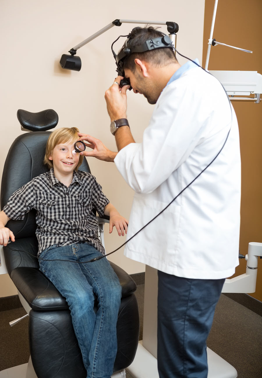
The patient will be asked to sit in a reclined position or to lie down on the couch. The doctor will wear a bright light, like a miner’s torch, on the forehead. He/she will shine the light in the patient's eye and hold an ophthalmoscope in front of the eye to help him/her observe it. The doctor can ask the patient to turn their eyes in specific directions as he/she sees the back of the eye. He/she might also use a small, blunt probe to put some pressure on the patient's eye.
The test allows the doctor to view the parts in the back of the patient’s eyes in greater detail. It includes the front parts of the retina, which are challenging to see with the other methods. The doctor may combine the exam with another technique called scleral depression to bring the far margins of the retina into view. It allows the doctor to see if the retina has tears or is detached.
Slit-lamp examination
The method gives the doctor a similar look of the patient's eyes as the indirect test and with higher magnification.
The patient will sit before an instrument called a slit-lamp. It has an area for the patient to rest their chin and forehead to maintain their head steady during the test. The doctor switches on a bright light on the patient's eye and uses a binocular microscope to examine the structure at the back of the eye. He/she might request the patient to roll the eyes in different sides, and open the eye with his/her fingers to have a better view. He/she can also use a little, blunt probe to exert a bit of pressure to the patient's eyeball through the skin of their eyelids. The pressure helps bring the fridges of the fundus into view.
The exam only takes a few minutes.
Outcome
The specialist will recommend treatment options or suggest further tests in case of abnormalities.
Abnormal results include:
- Swelling of the optic nerve
- Retinal tear or detachment
- Optic nerve damage due to glaucoma
- Clouding of the eye lens which may indicate a cataract
- An infection of the retina, Cytomegalovirus (CMV) retinitis
- A form of skin cancer that can spread to the eye (Melanoma)
- Bleeding in the back of the patient's eye or damaged blood vessels could point to chronic diseases such as diabetes and high blood pressure
- Presence of drusen, which are hard, yellow or white deposits under the retina, or leaking blood vessels (hemorrhages), may be an indication of macular degeneration. The age-related diseases cause a loss of vision in the center of the visual field.
Risks & Complications
In rare cases, the eye drops can have side effects including:
- Nausea
- Flushing
- Dizziness
- Vomiting
- Dry mouth
- Narrow-angle glaucoma
The patient should call the doctor immediately if they experience vision problems like halos around lights, loss of vision, or severe and sudden eye pain after the exam.
