Introduction
A gonioscopy is an eye exam used to evaluate the internal parts of the eyes. It can be part of the routine eye exam, depending on the age of the patient. The test aims at thoroughly checking the patient’s eye health and correctly diagnosing eye disorders such as glaucoma. It is among the leading causes of blindness. A gonioscopy combines a slit lamp and gonioscope to allow the doctor to examine the anterior chamber, the front area of the eye, which lies between the iris and the cornea. The exam can enable the doctor to evaluate internal structures that are not visible when using a penlight or slit lamp only.
Purpose
Gonioscopy test can be done at the doctor’s office or eye clinic by:
- An optometrist
- An ophthalmologist
The doctor can use the gonioscopy test to evaluate a patient with symptoms of glaucoma. The eyes continuously produce aqueous humor. It is the liquid substance that occupies the anterior chamber of the eyeball. So, fresh aqueous humor is always flowing into the central chamber of the eye. A tiny drainage duct at the corner of the eye allows the excess liquid to flow out. It works to maintain a stable internal eye pressure or intraocular pressure (IOP). Therefore, as flesh fluid flows into the chamber, a corresponding amount vacates through the drainage opening.
In a situation where the drainage system is abnormal or defective the fluid builds up in the chamber. For example, where there is a blockage of the drainage opening. This results in an increase in intraocular pressure, which can cause eye trauma or injury. A prolonged elevated IOP can result in optic nerve damage and the development of glaucoma. If not treated, the disorder can cause irreversible loss of vision or complete blindness. The doctor will conduct a gonioscopy to determine whether:
- There are congenital disorders that may eventually lead to glaucoma
- The drainage angle is open or closed. This identifies the type of glaucoma
The gonioscopic test also helps the doctor to observe other subtle details of the drainage system to guide diagnosis and plan appropriate treatment.
Often, it is recommended for patients with various conditions, including:
- Eye trauma
- Ocular hypertension
- Diabetic retinopathy
- Elevated eye pressure
- Narrow anterior chamber
- Pseudoexfoliation syndrome
- Central retinal vein occlusion
- Pigmentary dispersion syndrome
Gonioscopy, in combination with other eye tests is recommended for patients with the following symptoms:
- Double vision
- Excess tearing
- Change in iris color
- Abnormally dry eyes
- Seeing floating images
- Recurrent pain around the eyes
- Unusual sensitivity to light or glare
- Red-rimmed, swollen or encrusted eyelids
- Lines or edges appearing wavy or distorted
- Unexpected inability to adjust vision in dark rooms
Gonioscopy may be required in emergencies when the patient:
- Sees halos or rainbows around light
- Gets a sudden blurred or hazy vision
- Has flashes of light or black spots in vision
- Experiences a sudden loss of vision in one eye
Gonioscopy is also used along with other tests to examine and verify the gravity of eye abnormalities. The other tests may include:
- Slit-lamp examination
- Tonometry, to measure eye pressure
- Perimetry, to determine the range of vision
- Ophthalmoscopy, to check the optic nerves
Preparation & Expectation
The patient should inform the doctor if they have a family history or family member with glaucoma. Patients who use glasses or contact lenses will have to remove them before the test. If the patient wears contact lenses, they should bring glasses because they can’t wear the contacts for at least two hours after the exam. The patient may also have blurred vision and sensitivity to light for some time after the test. So the patient should bring someone along or arrange to be driven home after the test. There is no pain in a gonioscopy exam.
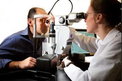
Procedure
The test requires the use of anaesthetic eye drops. The initial application of the medication may cause a slight burning sensation. The patient will rest their chin on a chin support and the forehead on a support bar. The doctor will place a special contact lens before the patient’s eye. He/she will then position the patient’s head in the slit lamp and shine a beam of light on the eye to illuminate the angle. The test only lasts a few minutes.
Outcome
In normal results, the drainage angle and canal will be open and sound. In abnormal results, the drainage angle will be narrow, blocked, or closed. This indicates the presence of glaucoma. Where the drainage angle remains open but there is a blockage inside the canal, it is known as open-angle glaucoma. It is the most common form of the disease and develops gradually without any pain. If the drainage angle is partially or wholly closed, it is called closed-angle glaucoma. The condition can develop slowly or suddenly. It is a severe form of the disorder and can lead to irreversible loss of vision.
The drainage angle can get blocked due to a variety of reasons such as:
- Eye injury or infection
- Abnormal blood vessels
- The presence of scar tissue
- Hyperpigmentation of the iris
After diagnosis, the doctor can recommend medication in the form of eye drops to lower the IOP and prevent or control the progress of glaucoma. In severe cases, the doctor can suggest surgery, such as trabeculectomy, to relieve the internal pressure. It allows the fluid to drain out of the anterior chamber and is the most frequently used glaucoma surgery.
Risks & Complications
There are some minor risks linked with the test, including eye infection and allergic reaction to the anaesthetic eye drops. It is critical for the patient not to rub their eyes or wear contact lenses until the eye drop medicine wears off. This prevents the scratching of the cornea, which can lead to an eye infection.
