Introduction
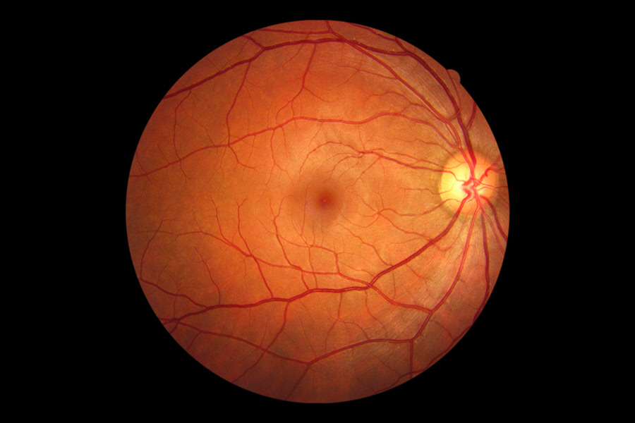
The fundus photo is an eye exam that uses a specialized camera (fundus camera) to capture images of the patient's fundus or inner lining. It is the region at the back of the eye comprising retina, vitreous, macula, choroid and the optic nerve. The fundus camera is an instrument resembling a microscope attached to a camera capable of taking pictures of the structures inside the eye.
Ocular fundus photography plays a vital role in monitoring the status of the patient's eye. The images help to document ocular abnormalities and disease progression. It is particularly useful in the diagnosis and management of retinal conditions such as glaucoma, macular degeneration, diabetic retinopathy, retinal hemorrhages, etc. It is also helpful for evaluating the success of retinal laser surgery. Fundus photography can also use specialized dyes and colored filters.
Also Known As
- Ocular fundus
- Fundus photo test
- Ocular fundus photography
Types
There are various forms of fundus photography, including:
- Fundus autofluorescence
- Standard fundus photography
- Smartphone fundus photography
- Ultra-widefield fundus photography
Purpose
The fundus photo test can be done at the eye clinic or the doctor's office by:
- A technician
- An optometrist
- An ophthalmologist
Fundus photos provide complementary information together with other eye tests such as perimetry and optical coherence tomography. It helps in the assessment of inner eye structures anomalies and in tracking the progression of diseases. It is critical in the observation of retinal disease processes such as:
Choroidal nevus
It is a melanocytic lesion of the choroid. It can have different sizes and pigmentation, though it frequently appears as a flat, dark and evenly pigmented lesion. However, choroidal nevi can also mimic other pigmented lesions of the retina, especially where a stereoscopic view is challenging to achieve. Fundus Autofluorescence is useful for differentiating these retinal lesions from melanomas. The test can be used to find out if a choroidal nevus has increased in size, which is a risk factor for melanoma.
Chorioretinal scar
Fundus imaging of chorioretinal injuries can help confirm whether there is a reactivation of lesions. It is challenging to describe chorioretinal injuries in words because they are irregularly shaped. Fundus imaging can establish the status of the wound (dormant or active) and detect lesions that may be invisible or challenging to notice with funduscopy. Photographic documentation provides a proper baseline and follow-up records, which enable the doctor to monitor the patient's condition.
Central serous chorioretinopathy
It is also called central serous retinopathy. Often, it is a self-limiting retinal disorder denoted by fluid build-up beneath the retinal pigment epithelium layer. It is suspected to be caused by prolonged emotional stress and steroid use. Fundus photos can show visual distortion and lingering pigmentary disturbances.
Diabetic retinopathy
Screenings for the condition have become common with an increase in the prevalence of diabetes worldwide. In many cases, no treatment is necessary, though it is vital to stratify the risk of progression to avoid the development of the disease. Fundus photos can be a great accessory in the management of these patients.
Glaucoma
Fundus photography plays a crucial role in the management of glaucoma. It enables the doctor to scrutinize the retinal nerve fiber layer and optic nerve head. The examination is an integral part of the assessment of a glaucoma suspect or patient. It is more accurate in the detection of disc hemorrhages than clinical study. Fundus photo documentation also provides a reliable baseline for monitoring the patient.
Age-related macular degeneration (AMD)
The central area of the retina can start to deteriorate with age. The patient may find it harder to focus and experience blurry vision. There are two types of AMD:
- Dry macular degeneration
The fundus photo will confirm the presence of drusen beneath the retina and atrophy. Drusen are waste deposits that can damage the macula and lead to loss of vision.
- Wet macular degeneration
Fundus imaging will show abnormal retinal blood vessels growing and fluid leakage. It will also show the pigment epithelial detachment associated with the subretinal fluid. Wet macular degeneration can cause a sudden loss of vision through the formation of scar tissue or bleeding.
Preparation & Expectation
The fundus photo test is a painless and straightforward process. If the patient wears contact lenses, they will have to remove them before the test. The doctor will dilate the patient's eyes before the pictures can be taken. It allows the device to make the best possible photos of the inner eye.
The medication will keep the patient's eye dilated for about 15 hours. During this time, the patient will experience increased sensitivity to light. So the patient should bring sunglasses and bring someone along or make arrangements to be driven home after the test.
The patients should tell the doctor if they are allergic to iodine and about any medications they are taking, including over the counter medicines and dietary supplements. The doctor will also need to know if the patient has glaucoma or a family member with the disease.
Procedure
The patient will sit facing the fundus camera and place their chin on the support and rest their forehead on the bar. The doctor will adjust the camera and take a picture of the patient's inner structures.
Outcome
In the case of abnormal results, the doctor may explain the treatment options or suggest additional tests for a more definitive diagnosis.
Abnormal results may indicate:
- Retinal toxicity
- Melanoma – cancer in the eye
- Anomalies of the central nervous system
- Glaucoma damage to the optic nerve head and nerve fiber layer
- Disorders of the choroid layer, such cysts, ciliary body atrophy, ciliary body detachments, etc
- Macular degeneration - Drusen under the retina and atrophy. There may also be abnormal retinal blood vessels, bleeding or scar tissue
Risks & Complications
The dilation medication may have an allergic reaction, including:
- Hives
- Nausea
- Sneezing
- Vomiting
- Dry mouth
- Increased salivation
- Increased heart rate
The doctor may treat the allergic reaction with shots or pills. In sporadic cases, the patient can have a severe allergic reaction, such as fainting, difficulty breathing, narrow-angle glaucoma, etc.
