Introduction
Corneal topography is a computer-assisted imaging method that produces a color-coded graph of the curvature of the cornea. The cornea is the transparent front section of the eye that works like a windscreen. It also functions as the primary lens by transmitting and focusing light on the back of the eye. Corneal topography provides detailed information about the form and power of the cornea.
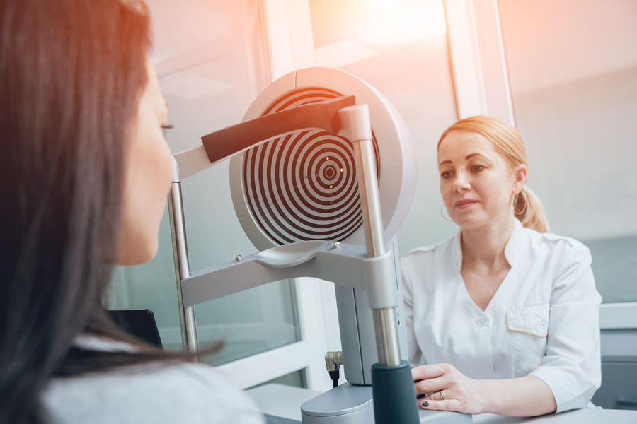
Corneal mapping uses computerized analysis and a specialized camera. It is a non-contact evaluation technique that photographs the cornea surface using ordinary light. It works by sending illuminated rings onto the surface of the cornea, which bounce back and are then measured by the device. The computer then creates a topographical image of the cornea. Corneal topography can help the doctor to detect abnormalities that are invisible when using conventional testing methods.
Also Known As
- Corneal mapping
- Videokeratography
Purpose
Corneal topography can be done at the doctor's office or eye clinic by:
- A technician
- An optometrist
- An ophthalmologist
Corneal mapping is not a routine test. The doctor can order it in exceptional cases to diagnose specific types of abnormalities, assess the progression of a disease, to fit particular types of contact lenses, and in the preparation of surgical procedures.
The exam can help the doctor to determine the curvature of the cornea and detect distortions like keratoconus. It is a degenerative disease that leads to a thinning of the cornea. The condition may be diagnosed with the initial exam. However, follow up tests may be necessary to assess whether the corneal surface is becoming more irregular or steeper with time.
The map can also assist the doctor in the evaluation, treatment and management of various corneal curvature anomalies and ocular conditions, including:
- Corneal opacities or scars
- Corneal deformities
- Corneal scratches
- Corneal diseases
Corneal mapping creates a three-dimensional map that is used in the planning of surgical procedures. The evaluation is useful for:
- Fitting contact lenses
- Corneal transplants
- Planning vision correction surgery
- Irregular astigmatism after corneal transplantation
- Postoperative cataract extraction with acquired astigmatism
Preparation & Expectation
The doctor may instruct the patient to stop wearing contact lenses for some time before undergoing the test. The period can range from a few days to several weeks. It helps to rule out temporal alterations on the shape of the cornea caused by wearing contact lenses. It also helps to improve the accuracy of the readings. So patients who wear contact lenses will do well to check with the doctor for guidance. The exam is brief and painless.
Procedure
Corneal mapping is a non-contact technique. The test is like taking a picture of the eye. If the patient has dry eyes, the doctor may apply some moistening eye drops to improve the quality of the photograph.
The patient will sit on a chair or stool and rest their chin and forehead on support. The doctor will instruct the patient to look at a light source or another target in the bowl of the mapping camera. They will be asked to keep their eyes open for several seconds as the image is captured. Once the picture is obtained, the patient can blink as usual as the computer produces a map of the corneal curvature.
The doctor may have to repeat the process several times before getting a high-quality photograph. He/she may review the image with the patient after the test and on subsequent visits.
Outcome
The cornea accounts for close to 70 percent of the refractive power of the eye. Therefore, its shape determines the visual ability of the eye. Corneal mapping results provide the doctor with comprehensive information about the shape. A healthy eye shows an evenly rounded cornea. Abnormal results may reveal a corneal curvature that is too flat, too steep, or uneven.
The doctor will use the results of the test to ascertain whether the patient has keratoconus. He/she will also be able to determine whether the disease is at a mild, moderate, or severe stage. Follow up measurements will provide information on whether the disease is progressing.
The doctor can also use the corneal mapping results to determine the best vision correction options and as a guide in contact lenses fitting. The results are frequently used in the planning for LASIK vision correction refractive surgery. The 3D map, together with results from other tests, can assist the eye surgeon to ascertain in precise terms the amount of corneal tissue that has to be cut out to correct the refractive error. It can also help the doctor to assess if the patient will benefit from corneal cross-linking.
Risks & Complications
Corneal topography is a noninvasive technique without any complications or risks.
