Introduction
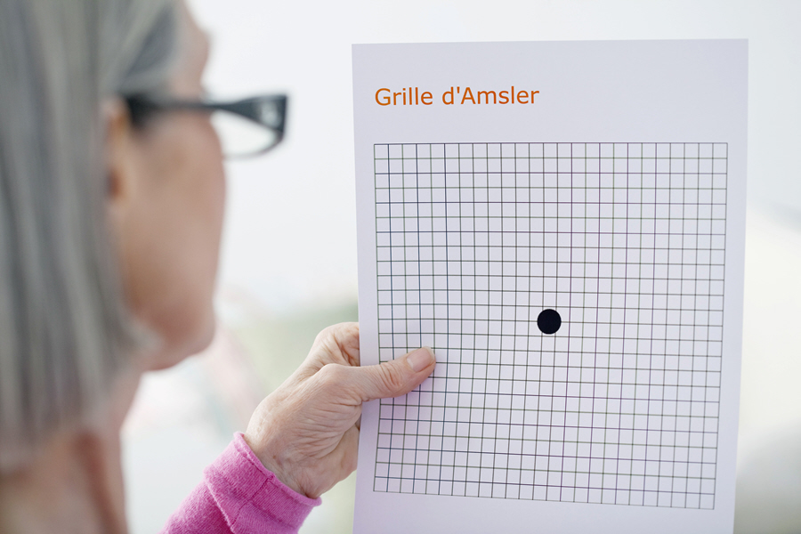
The Amsler grid is an eye chart consisting of vertical and horizontal lines. It resembles a piece of graph paper with a small spot or dot in the middle and helps to track a patient's central visual field. The eye chart was developed by a Swiss ophthalmologist called Marc Amsler and has been in use since 1945. It is an assessment tool that assists eye professionals in detecting visual abnormalities arising from changes in the back of the eye, the pituitary gland, the optic nerve, and the optical channel to the brain.
The original version was a black and white grid. Later, a blue and yellow chart and other color models were introduced. The blue and yellow chart is more sensitive and is useful for evaluating a broad range of visual channel disorders. The patient uses one eye at a time to look at the small dot in the middle of the table.
The Amsler grid is available from eye doctors or web sites.
Also Known As
Amsler chart
Purpose
The Amsler grid test can be done at the eye clinic, doctor’s office by:
- An optometrist
- An ophthalmologist
The patient can also conduct the test at home.
The doctor uses the Amsler grid to identify early warnings of retinal disease and track changes in the patient’s vision after diagnosis. The chart is particularly useful in the detection of macular diseases such as Epiretinal membrane anomalies and age-related macular degeneration (AMD). The doctor uses the grid to monitor patients with dry AMD. It helps the specialist to detect the progression of the disease to the wet stage early enough. Wet AMD may be treatable during the early stages.
The grid can also help the doctor to detect various disorders, including:
- Optic neuritis- an inflammation of the optic nerve
- Nutritional optic neuropathy- a disease where toxic reaction in the optic nerve causes a loss of vision
- Metamorphopsia- a form of distorted vision that makes straight lines in the chart appear wavy and areas of the table seem blank
- Scotomas- a patch of a partial change in the field of vision. It consists of partially diminished or completely degenerated visual acuity surrounded by a sphere of relatively well preserved vision
Preparation & Expectation
There is no special preparation required for the Amsler grid test.
Procedure
The patient will sit comfortably in an examining chair in a room with normal illumination. They will have the proper near correction aid, for example, reading glasses. Usually, the patient's spectacles are satisfactory. However, they should be single vision lenses or trial glasses and not bifocals.
The doctor will instruct the patient to hold the grid at about 30 centimeters. The standard grid is a white chart with five millimeters (5mm) squares on a black background. The table has a white fixation target at the center. The doctor may also use various versions of the table depending on what s/he is investigating. For example, s/he can use a table with random white dots and a white fixation target in the middle to check for scotomas. The standard chart with a red grid may be used to assess optic neuritis and nutritional optic neuropathy (toxic amblyopia). It can also help to expose the malingerer when combined with green and red filters.
The test begins with the patient holding the standard chart. The doctor will ask the patient to look at the table with one eye at a time (monocularly). S/he will want to know whether the patient can see the dot in the middle. It helps to ascertain if the patient has an absolute or central relative scotoma. The doctor will then instruct the patient to keep focusing on the middle spot for the duration of the test. However, the patient ought to be aware of the whole chart from the edges of their eyes.
During the test, the doctor will ask various questions, such as if:
- the patient can see all corners and sides of the large square/chart
- any of the squares in the table are missing or blurred
- any of the lines of the grid appear distorted or wavy
The doctor will note any disturbances or anomalies on an Amsler recording sheet. S/he may ask the patient to draw the abnormalities on a recording chart.
Home test
The patient can obtain a chart from the doctor or online. They should wear their reading glasses and hold the table at a distance that enables them to focus on the lines, say at about the reading distance. One should cover or close one eye and test each eye at a time. Otherwise, the healthy eye will compensate for the one with a problem, e.g., wet AMD, and the patient won’t notice any anomalies.
It is essential to note that the Amsler grid test does not replace a comprehensive eye exam.
Outcome
Often, to a patient with macular disease, some lines may seem to be missing or wavy. For instance, to an eye with wet AMD, some of the tracks will appear curved or even obstructed by a white, gray, or black patch. It is caused by the fluid that builds up under or within the retina. It can create a blister causing straight lines to appear curved. At times, the fluid can interfere with retinal function by creating a black, white, red, or a gray area near or in the center of the patient’s visual field. If wet AMD goes untreated for several months, it can develop retinal scarring leading to a permanent vision loss in some areas of the visual field.
A patient should use the grid to monitor their vision at home at least once a week. They should call the specialist if they notice any change. Even without a chart, an individual who sees changes in their vision should urgently call the doctor. It is mainly the case if:
- Reading becomes more difficult
- It becomes harder to recognize objects or faces
- Computer and TV pictures are more challenging to see
- Straight lines look curved, for example, a door or window frame
Risks & Complications
The Amsler grid test has no risks or complications.
