Introduction
Intermittent or subacute angle closure is a condition of the eye where there is spontaneous and recurrent ‘narrowing’ of the anterior chamber angle. The closure results in build up of internal pressure in the eye, what is referred to as intraocular pressure (IOP).
Angle closure occurs in three main forms; the acute form, the subacute/ intermittent form and the chronic form. Whereas the angle closure presents sudden dramatic symptoms, the chronic form is asymptomatic and is usually detected when performing other ophthalmic examinations. The subacute form presents symptoms similar to those of the acute but on a milder scale, with the episodes resolving spontaneously. All forms of angle closure can gradually lead to glaucoma and blindness.
Also Known As
Sub-Types
Causes and Risk Factors
Angle closure occurs when the peripheral iris gets into contact with the trabecular meshwork. This can be caused by a number of factors such as:
- Pupillary blockage - The back of the iris may adhere to the lens, blocking the pupillary and resulting in fluid back up. This fluid pushes the iris forward to the trabecular meshwork, eventually blocking the angle.
- Anatomic changes such as increased iris thickness, increased lens thickness or displacement of the iris in the case of plateau iris syndrome.
- Underlying eye conditions - Angle closure can also arise secondary to other eye diseases such as phacomorphic glaucoma, neovascularization or ectopic lens.
- Drugs - Some medications have also been shown to increase the risk of angle narrowing. These include pupil dilators and sulfa-containing drugs among others.
Risk factors for angle closure include old age, the female gender, being of the Asian or Inuit descent, and having hyperopia or a family history of angle closure.
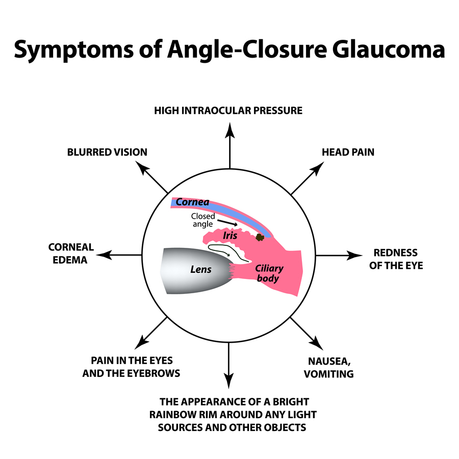
Signs & Symptoms
The signs and symptoms to watch out for include:
- Blurry vision with halos around lights
- Mild to moderate headaches
- Brow aches
- Nausea
- Vomiting
- Redness of the eyes
- Increased IOP
- Edematous cornea
Diagnosis
After presenting the symptoms to the eye professional, a number of tests can be performed. A comprehensive eye exam will include the following:
- Tonometry - Tells where there’s high IOP, that is levels above 21 mmHg
- Gonioscopy - Considered the definitive test as it shows whether the angle is closed or open
- Slit lamp examination
- Automatic static perimetry
Treatment
Treating intermittent angle closure involves medical or surgical interventions that aims to reduce the IOP, widen the angle or prevent optic nerve damage.
Medical Treatment
Glaucoma medications are commonly issued to lower the intraocular pressure in angle closure. Topical and systemic drugs administered include beta blockers and carbonic anhydrase inhibitors.
Surgical Treatment
The most common surgical operation carried out is the laser iridotomy. This is where a hole is made in the iris to act as an alternative channel for the aqueous humor, creating a regular flow of the fluid.
Besides that, extraction of the cataract lens is another approach to managing angle closure. It lowers the IOP in the eyes, preventing optic nerve damage.
Prognosis/Long-term outlook
With early detection, intermittent angle closure is easily manageable and the angle reversible. However, delayed treatment can result in the chronic form, damage to the optic nerves (glaucoma) and ultimately, loss of vision.
Prevention/Follow Up
Regular eye screening, particularly for those at a higher risk of developing the condition, is a preventive measure for angle closure. Those already diagnosed should also go for check ups as frequently as once per week during the treatment period until the IOP control is achieved.
