Introduction
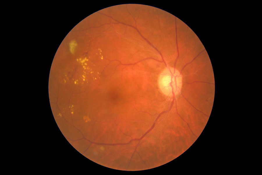
Macular edema occurs when fluid from damaged blood vessels builds up in the macula inside the retina. The retina is the area at the back of the eye that receives light and sends images to the brain. The macula is found at the center of the retina. With the macula, an individual can see details of an object in front of them such as written text.
The buildup of fluid in the retina can cause swelling leading to distorted vision. If macular edema is not treated, it can lead to permanent loss of vision.
Causes and Risk Factors
Abnormal leakage and accumulation of fluid in the macula causes macular edema. Several conditions can lead to this. They include:
- Age-related macular degeneration (AMD) – Abnormal blood vessels develop from the choroid and extend into the retina. The growth leads to leaking which causes swelling in the macula
- Diabetic retinopathy – The disease causes blood vessels to leak into the macula
- Retinal vein occlusion (RVO) – It occurs when veins in the retina get blocked such that fluid leaks into the macula
- Macular pucker/vitreomacular traction – When vitreous in an old person's eye fails to detach entirely from the macula, the vitreous forms scar tissue and tugs on the macula. The result is fluid collection underneath the macula
- Eye tumors – Whether malignant or benign, tumors can cause the macula to swell
- Inflammatory eye diseases – One such disease is uveitis which can cause harm to retinal blood vessels
- Hereditary disorder – They include retinitis pigmentosa (breakdown and loss of cells in the retina)
- Trauma to the eye – Objects like twigs may injure the eye causing fluids to leak
- Eye surgery – For example, cataract surgery. Macular edema develops a few weeks after surgery
- Medication – Certain medicines can lead to macular edema such as topiramate which is used in the treatment of seizures
Signs & Symptoms
In the beginning, macular edema does not present any symptoms. However, once the blood vessels start leaking, the following may be evident:
- Colors seem different or faded
- Difficulty reading
- Blurry or wavy central vision
- Noticeable vision loss
- Lack of pain in the eye
Diagnosis
The eye care professional will do the following to diagnose macular edema:
- Visual acuity test to identify vision loss
- A dilated examination to see the back of the eye where the retina is located
- Amsler grid test to check for any changes in the central vision; it recognizes even the smallest changes in one’s vision
- Fluorescein coherence tomography where the professional injects a yellow dye into the arm. The dye moves through the bloodstream to the retina. He/she then takes photos of the retina using a special camera as the dye travels. This test can reveal the leakage as well as its intensity.
- Optical coherence tomography (OCT) - It involves a machine scanning the retina to provide detailed images of its thickness. The test assists in measuring the edema.
Treatment
Treatment of macular edema aims to treat the underlying cause of the condition.
Medical Treatment
Treating the diseases or conditions that cause macular edema will address the problem. They may include:
- Inflammation – Using steroids to treat inflammation. Medications include pills, injections or eye drops. Examples of these medicines are ozurdex, retisert, Iluvien and so on.
- Cystoid macular edema after cataract surgery – Patients can use Non-steroidal anti-inflammatory (NSAID) eye drops for a few months. They can also use NSAIDs before and after cataract surgery to prevent macular edema from developing. They are also necessary when the eye is not responding well to steroids or to avoid side effects of steroids.
- Abnormal blood vessels in the retina – Anti-VEGF medication can reduce abnormal blood vessels in the retina. They also may lessen leaking from blood vessels. The eye doctor will use a very slender needle to inject the medicine into the eye.
- Glaucoma – Glaucoma medications include steroids and topical medications.
Surgical Treatment
- Laser surgery stabilizes vision. The surgeon uses laser pulses to seal off leaking blood vessels in areas where the fluid is leaking around the macula.
- Vitrectomy surgery restores the macula to its normal position after the vitreous has pulled away. To relieve the traction damaging the macula, the surgeon removes the vitreous from the eye and peel scar tissue from the macula. Vitrectomy also corrects vision when other treatments for macular edema fail.
Prognosis/Long-term outlook
Macular edema may take several months to clear depending on the cause and recommended treatment. The patient must strictly adhere to the treatment for it to be effective.
Macula edema that is caused by eye surgery often responds well to treatment because it is mild.


