Introduction
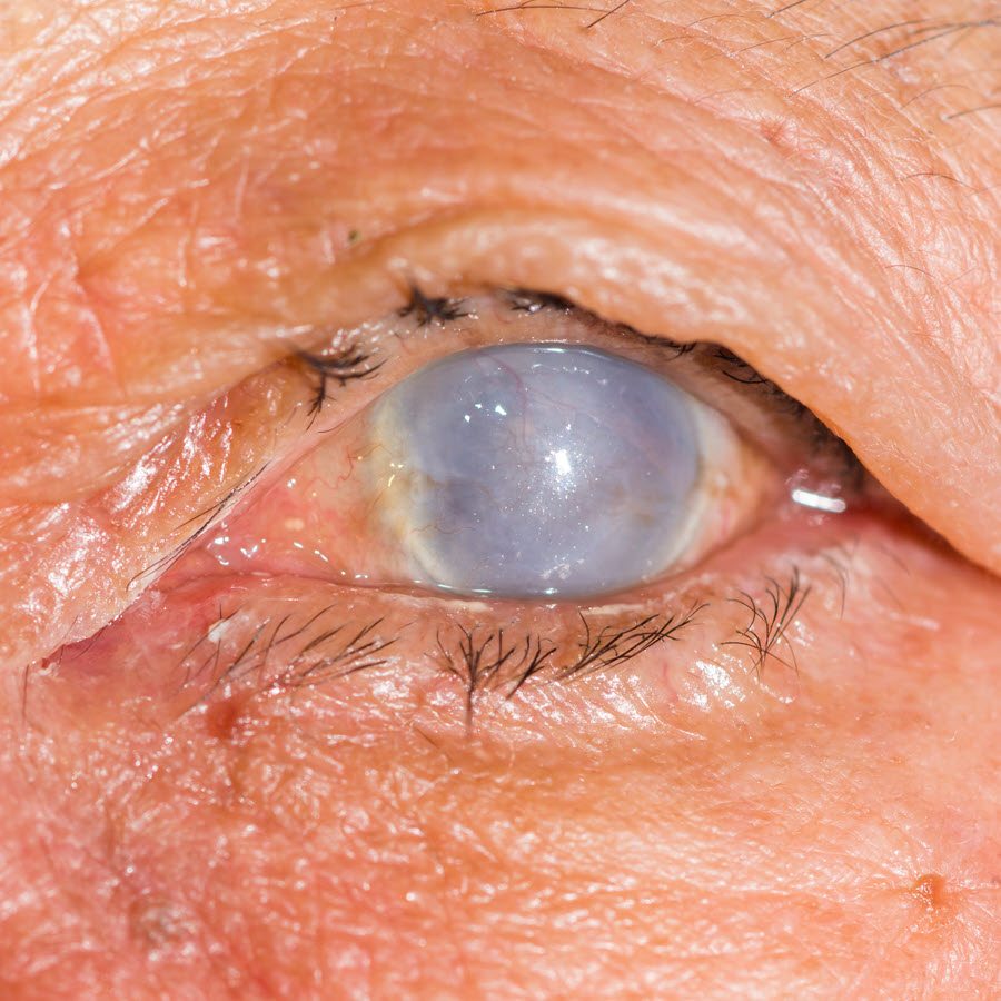
Corneal opacity is an eye disorder that causes scarring of the cornea. It is responsible for significantly decreased vision in affected individuals.
The cornea is the transparent part on the front of the eye. It protects the eye from dust, direct UV rays among other harmful foreign substances. Besides that, it allows light to pass through it before focusing on the retina. When scarring occurs, light is not refracted as it should be. Due to this, images are not correctly focused and vision is distorted in affected persons. The opacity may be located at the centre, at the periphery or may cover the whole cornea (opaque).
Also Known As
- Congenital corneal opacity
- Acquired corneal opacity
Causes and Risk Factors
Corneal opacities can be congenital or acquired. Congenital corneal opacities are present from birth. They are said to be caused by a malformation of the anterior segment of the eye. However, other causes can be congenital glaucoma, infections, metabolic storage diseases or trauma.
Acquired opacities, on the other hand, commonly arise from infections. Keratitis, associated with an infection, is a condition known to cause opacities. It is characterized by inflammation. Injuries are also responsible for some corneal opacity cases. Corneal abrasion is a common injury associated with opacities.
Risk factors for opacities may include:
- History of injury
- Wearing contact lenses for long periods - This can increase chances of eye infections and thereby the risk of developing opacity
- Measles - Can cause scarring of the eye
- Having infections - Infections such as Herpes simplex virus can be transmitted to the eyes. Conjunctivitis also increases the risk of acquiring corneal opacities
- Deficiency of Vitamin A - Vitamin A in the body reduces the risk of infections. A deficiency in the vitamin increases risk of infections and can result in opacities.
- Abnormalities in the cornea - Infants born with central corneal opacities are likely to have corneal abnormalities.
Signs & Symptoms
The symptoms will depend on the cause. However, patients may experience the following:
- Decreased or complete loss of vision
- Cloudy area on cornea
- Pain
- Foreign body sensation (Feeling as if there’s something in the eye)
- Redness
- Watery eyes
- Sensitivity to light
Diagnosis
The tests that may be carried out for diagnosis of corneal opacities include the following:
- Dilated eye exam - Drops are applied in the eye to dilate the pupil. The intention is to be able to view the inside of the eye properly.
- Slit lamp test - Through the use of a powerful and specialized microscope, the doctor can focus light into the eye for assessment. The slit lamp is used to examine the cornea and other parts of the eye for any defects.
- Visual acuity test - The test assesses vision of the patient. Defects should raise an alarm for opacities.
- Fluorescein staining - Staining is used to assess for any abrasions/scratches that may have caused the opacity.
Treatment
Treatment is aimed at preventing further damage to the cornea. It also focuses on preserving vision.
Medical Treatment
The options for medical therapy include:
- Eye drops - They may contain antibiotics to prevent infections. Doctors can also prescribe steroidal eye drops to ease inflammation and reduce scarring.
- Oral medications
Surgical Treatment
- Phototherapeutic keratectomy - This is a type of laser surgery. It is effective in the removal of corneal opacities among other ocular irregularities.
- Optical Iridectomy - For central corneal opacities which do not affect the entire cornea, the iris alone can be cut instead of the whole cornea.
- Corneal transplant - In the case of a severely damaged cornea (opaque corneal opacity), a transplant can be carried out. Patients should talk to their doctors about their medications before undergoing the surgery. Some drugs may need to be stopped for a specified period before the procedure.
Prognosis/Long-term outlook
Corneal opacities can easily lead to loss of vision. Prognosis for those with secondary corneal opacities is also poor due to the primary condition. If treated though, vision can be preserved.
Prevention/Follow Up
Preventive measures that can be taken to reduce the risk of developing opacities include:
- Wearing protective eyewear when in a potentially dangerous environment to avoid injuries.
- Monitoring vision and going for eye check ups in case of infection or injury
- Proper usage of contact lens
