Introduction
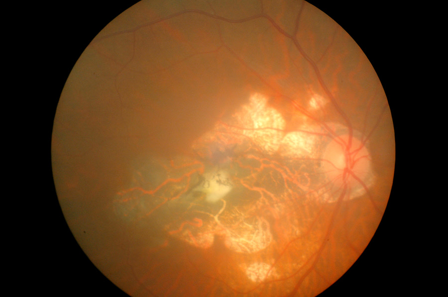
Choroidal neovascular membranes (CNVM) refers to new, destructive blood vessels which develop underneath the retina. These blood vessels grow in the choroid. The choroid is the part of the eye that is located between the retina and sclera. It forms part of the uvea and consists of connective tissue and blood vessels. Between the retina and choroid is the Bruch’s membrane, a thin layer of tissue. CNVM occurs when the blood vessels break through the membrane causing loss of vision.
The eye structures affected are usually those which give central vision to enable an individual to read, drive and even recognize faces. However, peripheral vision is not affected. This means an individual cannot become totally blind.
Also Known As
- Subretinal neovascular membrane
- Choroidal neovascularization
- CNVM
Sub-Types
- Type 1 - The neovascular membrane is found below the retinal pigment epithelium (RPE)
- Type 2 - The neovascular membrane passes through the RPE and lies above the RPE in the subretinal area
- Type 3 - Retinal angiomatous proliferation (RAP) which develops within the neurosensory retina
Causes and Risk Factors
A definitive cause of CNVM remains unknown. The disease is, however, associated with certain serious eye diseases which include:
- Wet age-related macular degeneration – Damage on the macula in the retina
- Histoplasmosis – An infection caused by the fungi histoplasmosis
- Eye injuries – Caused by sports, activities, foreign bodies and so on
- Myopic macular degeneration - A type of macular degeneration that occurs in people with severe myopia (shortsightedness)
- Angioid streaks – It’s associated with sickle cell anemia
CNVM is popular with people aged 50 and above. However, CNVM may occur in younger people if they have certain eye diseases or have an injury. The condition is also more common among Asian people.
Signs & Symptoms
The symptoms of CNVM may include:
Painless loss of vision in one eye
- Blank spots on one’s vision
- Distortion of vision like straight lines appearing crooked, irregular or bent
- Colors looking different for each eye or no longer appearing bright
- Each eye recording a different size of an object
- Flashes of light appearing in the central vision
Diagnosis
The eye care professional will do the following to diagnose CNVM:
- Conduct an eye examination by the use of a slit lamp, biomicroscopy and a fundus contact lens.
- Use fluorescein angiography to take a picture of the eye. The professional will inject a fluorescein dye into the individual’s arm. As the dye passes through the blood vessels in the retina, the eye doctor will take photos. The dye will highlight any abnormal areas enabling the him/her to diagnose CNVM.
- Use optical coherence tomography (OCT) to scan the eye. OCT creates a cross-section image of the retina that shows if there are abnormal blood vessels.
Treatment
Treatment of CNVM aims to eliminate the growth of abnormal blood vessels in the retina.
Medical Treatment
One way of treating CNVM is by use of anti-VEGF medications. Anti-VEGF drugs work against a chemical in the body that is responsible for the abnormal growth of blood vessels beneath the retina. The chemical is called VEGF or vascular endothelial growth factor. The drug slows down the leakage of the blood vessels. This action may slow the loss of vision. The eye doctor will inject the anti-VEGF drug straight into the eye in this outpatient procedure. He/she may administer anti-VEGF shots over many months for a prolonged benefit.
Surgical Treatment
The doctor may also use thermal laser therapy to treat CNVM. This procedure uses a high-energy focused laser beam. The laser produces a burn on the treatment area thus destroying the abnormal blood vessels. Surgery prevents more leakage, bleeding and growth of abnormal blood vessels.
Photodynamic therapy (PDT) is another surgical option used to treat CNVM. PDT is a combination of a special low-power laser and a photosensitizer (a light-activated drug). The surgeon will inject the medication into a vein in the patient's arm. Once the drug has moved to the abnormal blood vessels, the laser beam is focused on the vessels. This action activates the drug leading to the destruction of the undesirable blood vessels.
Prognosis/Long-term outlook
Treating CNVM helps to stabilize vision and checks further loss of vision. Lost vision cannot be recovered.
Laser usually causes a more blurred vision than was present before treatment. However, vision stabilizes in a few weeks. Laser treatment also causes a scar in the area the surgery occurred. The scar leaves a permanent blind spot which can be seen in the individual's field of vision. Although laser treatment destroys the abnormal blood vessels, a patient will need to repeat the procedure in three to five years.
Multiple treatments using PDT may be required because the abnormal blood vessels may recur.
Prevention/Follow Up
he following can help prevent CNVM:
- Individuals with a history or at risk of macular degeneration should get regular checkups
- The Amsler Grid should be used for monitoring purposes at home
- People over 60 years should have a comprehensive vision examination done annually
