Introduction
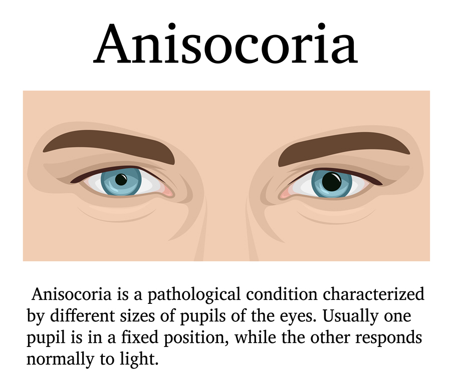
When there is a difference in the size of the pupils, an individual is said to be having anisocoria. The pupil is the black part in the centre of the iris. The iris is the black area of the eye. It controls the pupil to let in the right amount of light. When the light is deemed, the pupil widens (dilates). Bright light causes the pupil to narrow (constrict). The diameter difference in the size of the pupil can range from less than 0.5 mm to 1 mm.
One in five people have unequal pupil size with 20% of the population having physiologic anisocoria.
Genetics could predispose a baby to be born with anisocoria. It is nothing to worry about since the baby may not have any underlying disorder.
Sometimes pupils may differ in size on their own in adults without a cause. In such situations, there should be no cause for alarm especially when the pupil returns to normal. However, individuals who develop anisocoria later in life where pupils differ in 1 mm should seek treatment. It could be a sign of blood vessel, brain, nerve or eye disease.
Sub-Types
- Physiologic – When diameter difference is 1 mm or less, both pupils respond rapidly and uniformly to light
- Pathologic – It is caused by eye inflammation, trauma, eye medication, ciliary ganglion or nerve damage. The diameter difference is more than 1 mm in dim illumination and one pupil reacts poorly to light. Also, the pupil is shaped irregularly
Causes and Risk Factors
The following may cause anisocoria:
- Physiologic anisocoria is natural
- Horner syndrome which is sympathetic paresis affecting the eye. The other names for it are oculosympathetic paresis and Claude Bernard-Horner syndrome
- Third nerve (oculomotor nerve) palsy which may affect parasympathetic innervation to the pupil
- Diabetic oculomotor nerve palsy
- Eye injuries or trauma
- Head injury leading to bleeding inside the skull
- Medications such as homatropine (used to treat inflammation), asthma inhalers, eye drops and so on
- Substances that accidentally enter the eye such as insecticides
- Glaucoma that exerts excess pressure in one eye
- Inflammation of the iris
- Brain aneurysm
- Brain tumor
- Intracranial pressure caused by the swelling of the brain, acute stroke, intracranial tumor and intracranial hemorrhage
- Infection of membranes around the brain such as meningitis
- Migraines
- Seizures
- Cataract surgery
People most at risk of developing pathologic anisocoria include those with a nervous system disorder and stroke. They also include people with a history of damage to the eye, viral infection and Adie’s tonic pupil (one pupil has a sluggish response to light).
Signs & Symptoms
The signs of anisocoria may include:
- Horner syndrome – The presence of ptosis (drooping eyelid) on the side of the smaller pupil
- Third nerve palsy – Damage to the oculomotor nerve
- Irregular shape of the pupil (anterior uveitis, tonic pupil)
People with anisocoria may present the following symptoms:
- Problems with eye movement
- Headache
- Pain
- Reduced sweating
- Fever
- Double or blurred vision
- Vomiting or nausea
- Sensitivity to light
- Dizziness
- Cough
- Shortness of breath
Diagnosis
The eye professional will conduct an examination starting with a medical history. For example, they may want to know if the individual has always had pupils of unequal size. He/she may request to see an older photo of the individual.
The professional will check how the pupil responds to light in both a bright and dark room. He/she will also check the inside of the eye and how well the eyes focuses. The goal is to determine whether it’s the smaller or larger pupil that is the problem.
An eye drop test to see how the pupils react to medications that cause them to widen or narrow will also be conducted. Also, a slit lamp (instrument that uses high magnification) examination will be conducted.
The following tests may be done if there is need:
- A complete blood count (CBC) and blood differential tests
- Lumbar puncture (cerebrospinal fluid test)
- MRI and CT scan of the head (for people with Horner syndrome and third cranial nerve paralysis)
- CT scan of the chest (for people with Horner syndrome)
- Tonometry if glaucoma is suspected
- Electroencephalogram (EEG) test to measure the brain’s electrical activity
- A neck x-ray
Treatment
Treatment of anisocoria is directed towards correcting the underlying disorder.
Medical Treatment
Anisocoria itself does not need treatment since it does not affect eye health or eye sight. It is the underlying eye health problem that is treated. For example, if the anisocoria is caused by acute angle glaucoma, medication to decrease aqueous humor production may be prescribed.
If the patient has unequal accommodation, reading glasses or bifocals may be recommended. Patients affected by glare may use sunglasses or FL- 41 lenses.
Surgical Treatment
Surgery is dependent on the specific cause. For example, a surgical procedure to repair injured tissue caused by trauma may be done.
Prognosis/Long-term outlook
Follow up, prognosis and long-term outlook depend on the underlying diagnosis. For instance, Horner syndrome is life-threatening due to carotid dissection. Third cranial nerve palsy is equally life-threatening due to an aneurysm (weakening of an artery causing it to bulge). These are serious conditions requiring immediate medical attention.
